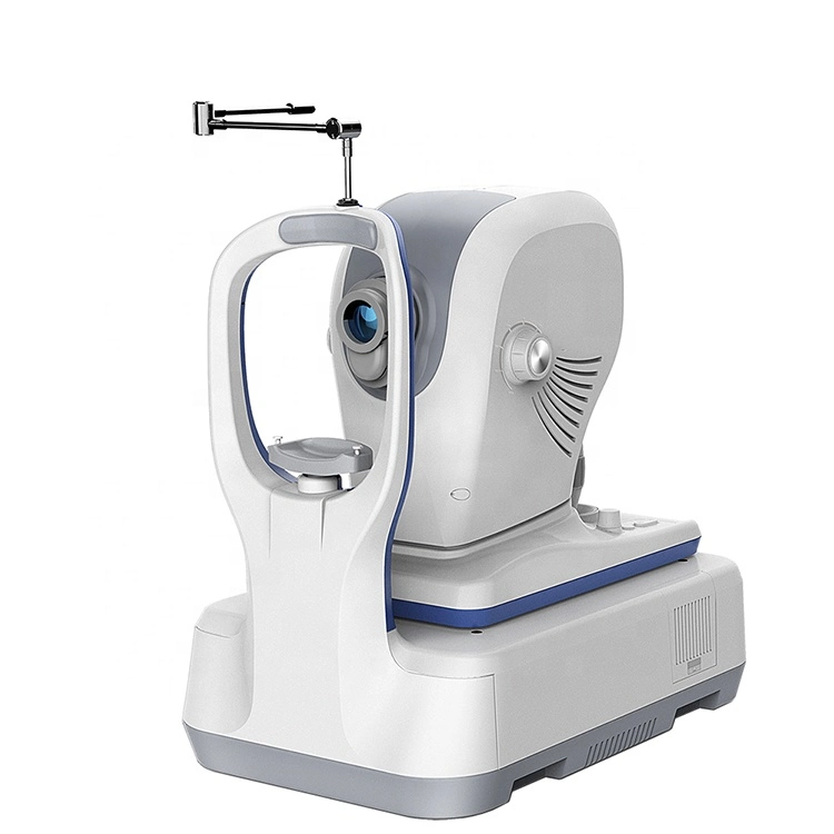
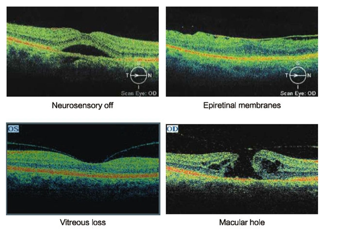
Ophthalmic Oct Equipment Optical Coherence Tomography Mce- Ose-2000

Min.Order: 1
Product origin: Guangzhou, Guangdong, China
Infringement complaint: complaintComplaint
US$ 1 ~ 999999

Description
Ophthalmic Oct equipment Optical Coherence Tomography
MCE- OSE-2000

Technical Data

.Optical coherence tomography (OCT) is a new , noninvasive , noncontact , transpupillary imaging,technology which can image retinal structures in vivo with a resolution of 5-8 microns. Cross-sectional images of the retina are produced using the optical backsattering of light in a fashion analogous to B-Scan ultrasonography and cofocal microscopy. Cross-sectional images of the retina, is revolutionizing the early detection and treatment and greatly enhanced our quality of patient care.
   Applications
In vivo,cross-sectional images and quantitative analysis of retinal fratures to optimize the diagosis and monitoring of retinal disease and for enhanced pre-and post-therapy assessment.
High-quality images and accurate measures RNFL and the optic nerve head to aid in the detection and management of glaucoma.
Cross-sectional images are valuable for clinical evaluation of macular holes, macular edema and other retinal pathologies. 
 
Precise location of pathology to expand disgnostic confidence and therapeutic precision.

Normal and abnormal image contrast image
Lesions image by image with normal OCT images, the regional thickness values? Topographic maps, diagrams and other multi-thickness contrast, thereby comprehensive judgment of disease.

High performance at a low price.
Modular design increases flexibility , reusability and maintainability.We can provide personalized design according to the customers needs.
With the powerful software , OSE-2000 has clear , easy-to-ure interface and supports multi-language.
 
 Anterior segment analysis template
Anterior segment with optional modules, OSE-2000 be able to observe and analyze the anterior segment.


TOMOGRAPHIC IMAGING
 
PURPOSECross-sectional imaging of the retina
SIGNAL TYPESuper luminescent LED, 840nm
LIGHT SOURCE≤0.75mW (Cornea)
OPTICAL POWER5μm in tissue
AXIAL RESOLUTION15μm in tissue
LATERAL RESOLUTIONGalvanometer mirror
SCAN MODEPosterior Segment: line scan, circular scan, cross hair scan, X-line scan, raster lines scan, radial lines scan, area scan
 Anterior Segment: line scan, radial lines scan
SCAN RAGE29,000 A-scans per second
ACQUISITION TIME58 pictures per second
SCAN DEPTH2μm in tissue
FUNDUS IMAGING 
SIGNAL TYPECCD imaging
FIELD ANGLE29°x23°
VIEWING METHOD22-inch color flat panel dispal
ILLUMINATORLED
INTERNAL FIXATIONLED dot matrix
EXTERNAL FIXATIONAdjustable blinking LED
MINIMUM PUPIL DIAMETER2mm

More ophthalmic equipments of our company


Product Tag:
Related categories:
Scroll to Top