 Complaint

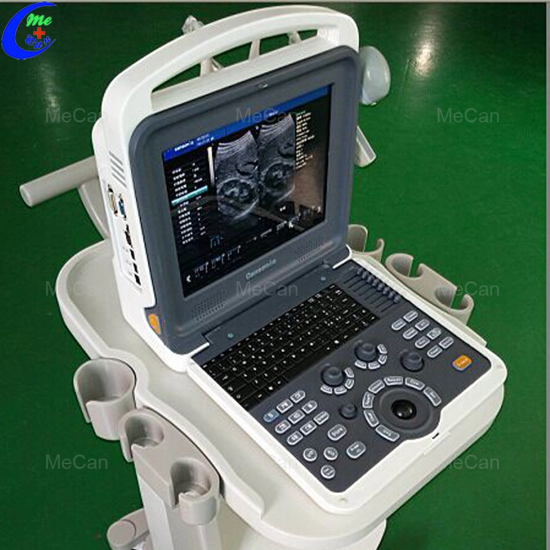
Model:MC-K2
What is the application of our 4d color doppler ultrasound?
Abdomen, Obstetrics, Gynecology, Pediatrics,Small parts, Artery, Superficial organ, Orthopedics,Cardiology, Musculoskeletal, Vascular, etc
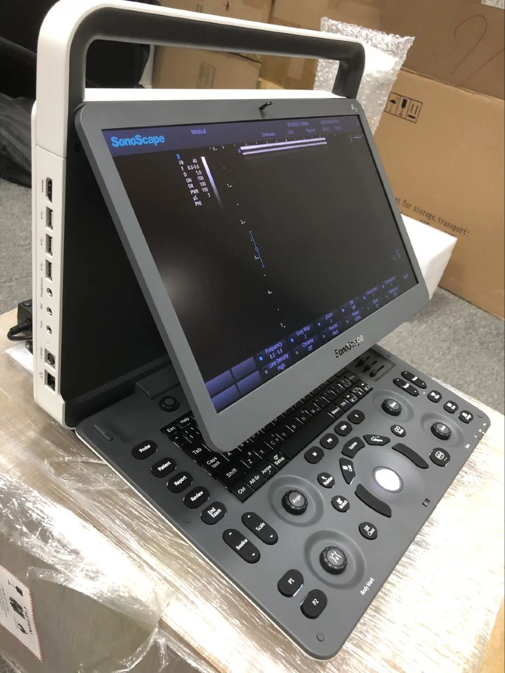
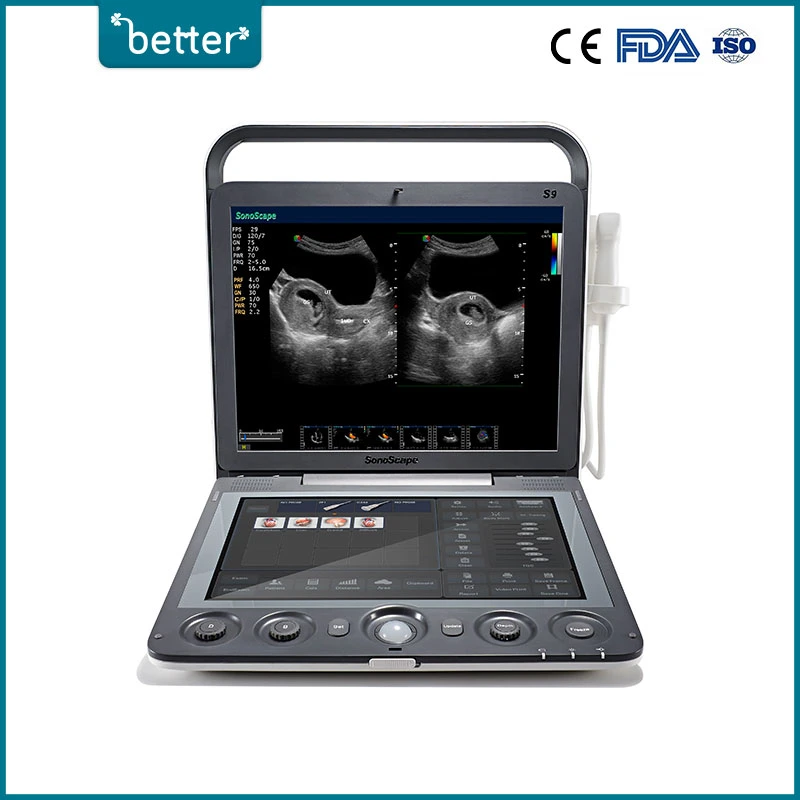
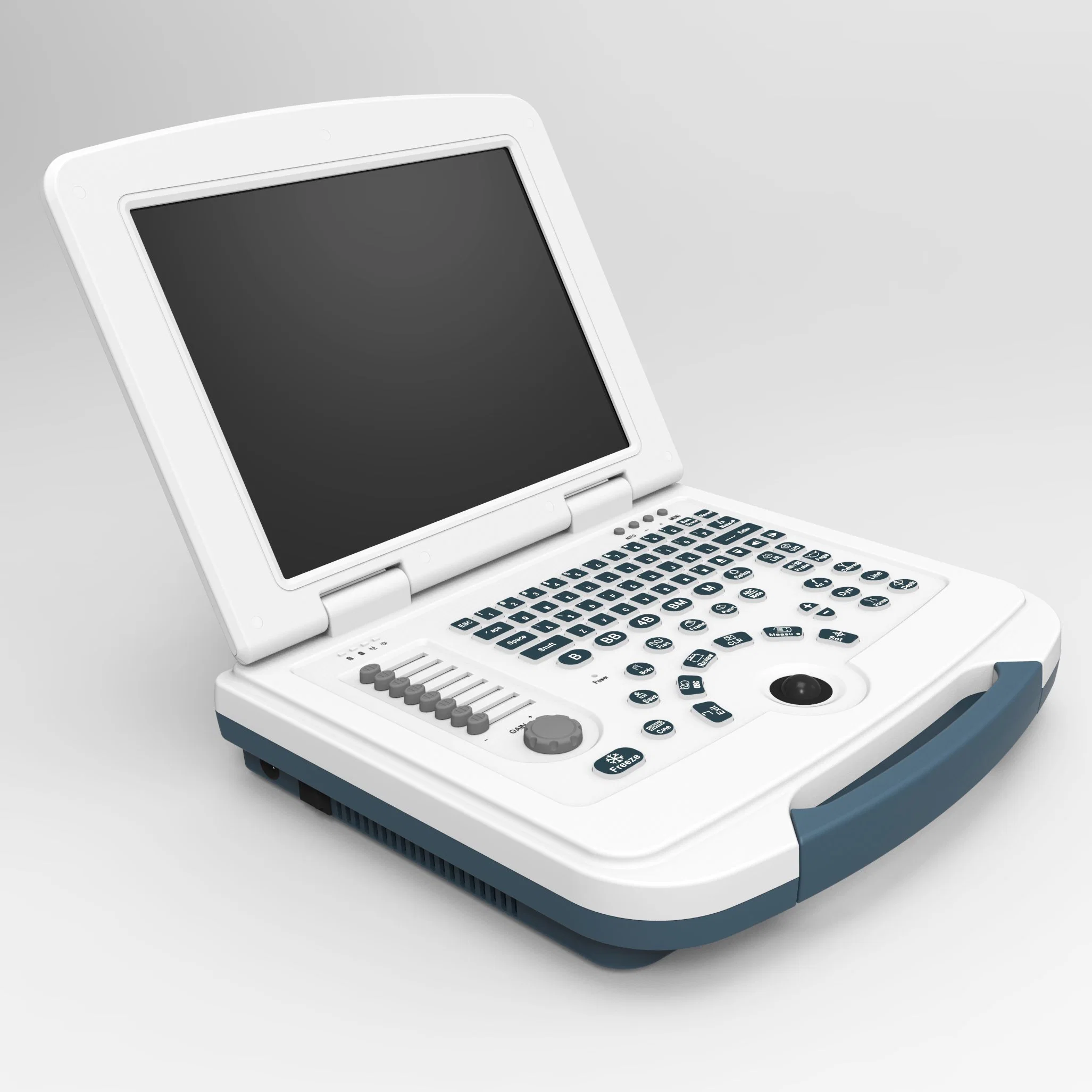
Smart, compact and clamshell design
12 inch LED monitor
Backlit operation panel, 8TGC
Floating keyboard
Two active probe connectors
Two probe holders
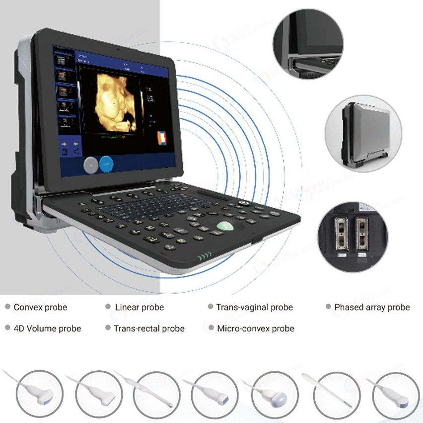
What is the specification of our echo machine?
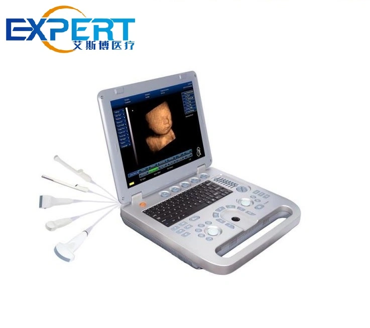
Transducer Types
Electronic convex probe
Electronic linear probe
Electronic micro convex probe
Electronic transvaginal probe
Electronic phased array probe
4D volume probe
Function
Auto Image Optimization
Tissue Harmonic imaging
iClear (Speckle Noise Reduction)
iBeam (Spatial Compound Image)
iZoom
PIHI (Pulse-Inverse Harmonics Imaging)
SA (Synthetic Aperture Synthesis)
Panoramic Image (Option)
Trapezoid Image (Option)
Continuous Wave Doppler(Option)
Display mode
B, B|B,4B, B|M,M,B|D,PW,B|PW,CW,CF
Triplex mode
4D mode
Zoom
Real time zooming
10 Steps: ×1.0, ×2.0, ×3.0, ×4.0, ×5.0, ×6.0, ×7.0, ×8.0, ×9.0, ×10.0
Selectable zooming position
Focus
Continuous dynamic focus
1~16 selectable transmit focus
Acoustic lens focus
1, 2, 3, 4 focus
Memory
Cine memory
B mode
M mode
SSD (Solid State Disk) 64G
Connectivity
Video out port
DVI out port
VGA out port
2 USB port
DICOM 3.0
Dimension
Gross dimension:510 mm X 500 mm X 330mm
Net dimension:330mm X 150 mm X 380mm
Weight
Gross weight:12 kg
Net weight:7 kg
Power Requirements
Voltage: AC 100V to 240V±10%
Frequency: 50Hz±1Hz
Rated Power: 250VA
Operation Conditions
Ambient temperature: 0ºCto +40ºC
Relative humidity: 38% to 85%
Atmospheric Pressure: 700hPa to 1060hPa
Standard Accessories
Power Cable
Operation Manual
Fuse
System Recovery USB
Built in Li-ion battery
Optional Accessories
B/W or color Video printer
LaserJet or inkjet printer
Trolley
Aluminum case
Biopsy guide
| B mode | 8 step TGC slide pots Gain: 0~100% Depth: 1.6~30cm Frequency: 5 steps Dynamic range adjustable: 0~150dB Edge enhancement:0~7 Persistence:0~7 Chroma:0~6 Grayscale:0~16 Power: 0~100% Noise reduction: 0-6 iclear: off, 1, 2, 3, 4 |
| M mode | Gain: 0~100% Sweep speed: 4 steps Maps: 0~16 Chroma:0~6 |
| C mode | Gain: 0~100% Pulse wave Wall filter: 4 steps Color Maps: 0~7 Package size: 8~15 Color persistence: 0~7 Threshold: 0-3 Base line: 0-6 Line density: Low and high Spatial filter: 0-3 |
| PW mode | Gain: 0~100% Frequency: 5 steps Pseudo color:0~6 PRFd:1.0~6KHz Basic line: 7 steps Wall filter: 7 steps Spectrum mode: Refresh and Synchronize Sampling volume: 0.5-48mm |
| B mode (General) | Distance Trace Length Ellipse (area) Trace(area) Angle Volume |
| PW mode | HR (heart rate) Distance Velocity Time |
| Abdomen | Liver Gallbladder Pancreas Spleen |
| Urology | Kidney Ureter Bladder After the urine bladder Prostate |
| Gynecology | Uterus Cervix Ovary Follicle |
| Early Obstetrics | GS BPD CRL NT |
| Later Obstetrics | BPD HC AC OFD FL TAD |
| Small parts | Thyroid Testes |
| Musculoskeletal | Hip |
| Peripheral vascular | Intima Artery |
| Cardiology | Distance Angle Volume RVWd LVDd RVDd LVPWd RVWs LVDs RVDs LVPWs RV/LV AO |
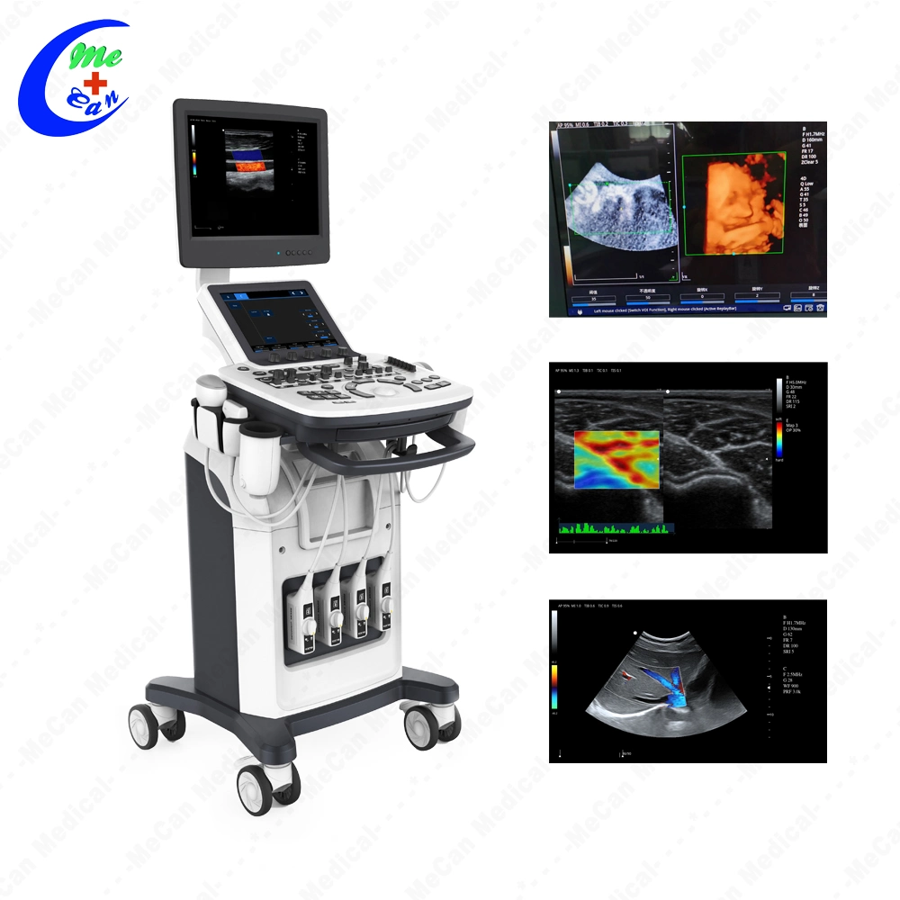
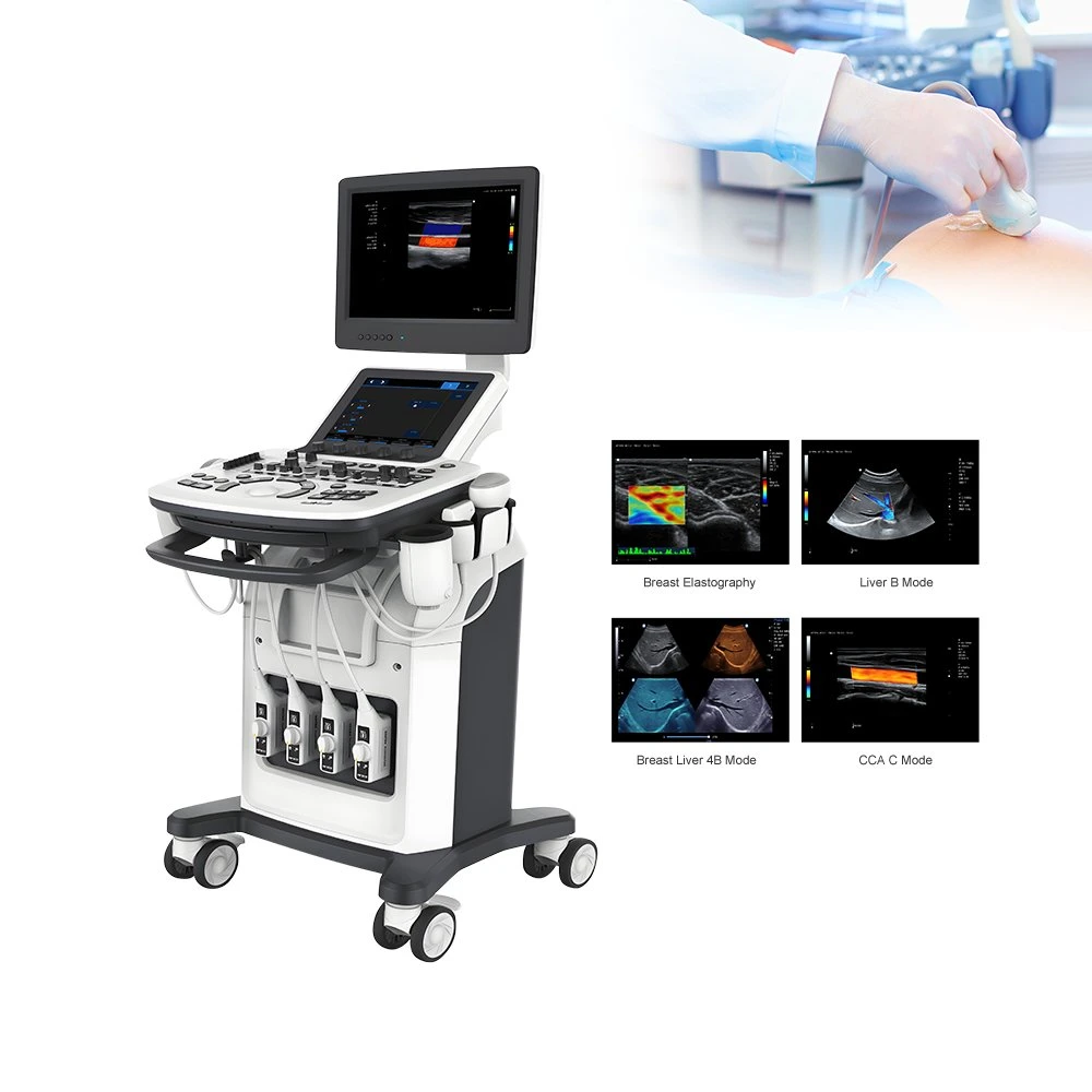
What is the Clinical Images of our echocardiography machine?