Description
Welcome to KS.Medcal
Guangzhou KuiShan Equipment Co., Ltd.
1.More than 5 years experience.
2. One-stop service.
3. The complete certificates and approved factory price.
4. Main Products: ECG, Ultrasound Machine, Laboratory Equipment, Ordinary Diagnosis Instrument, Patient Monitor, Operating Lamp and so on.
Product Details
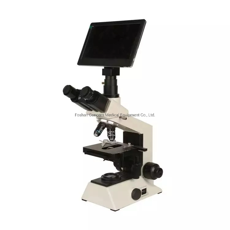
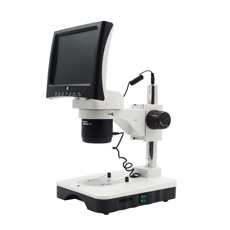
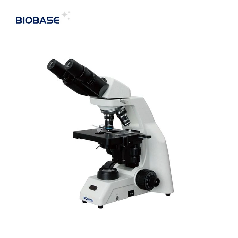
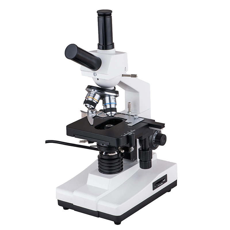
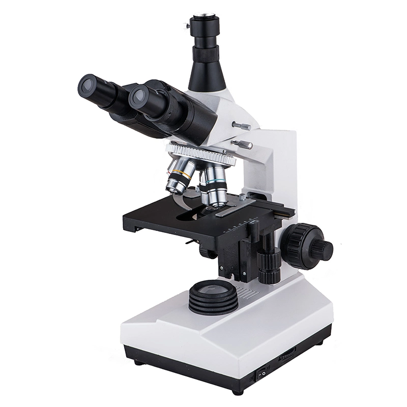
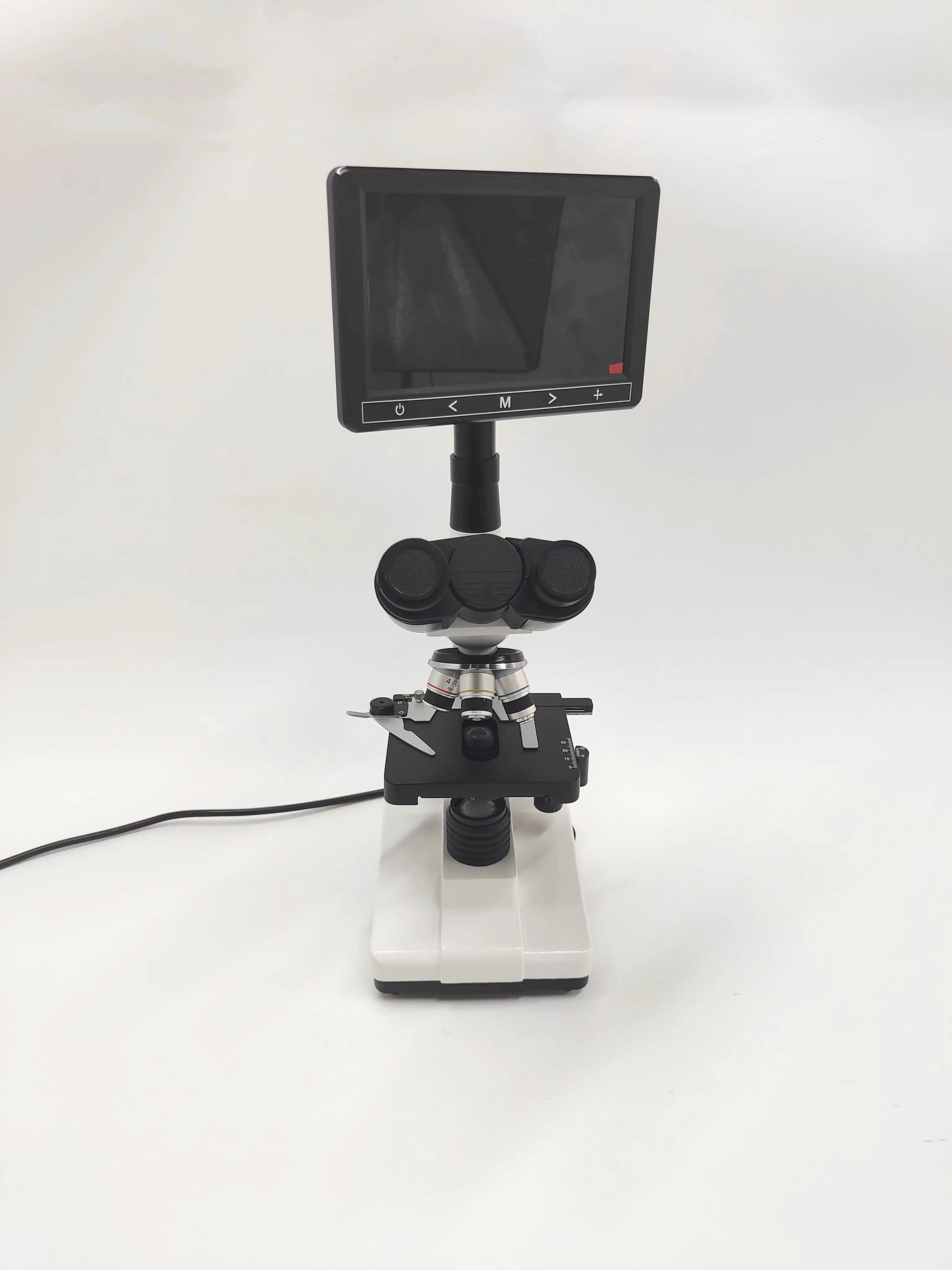
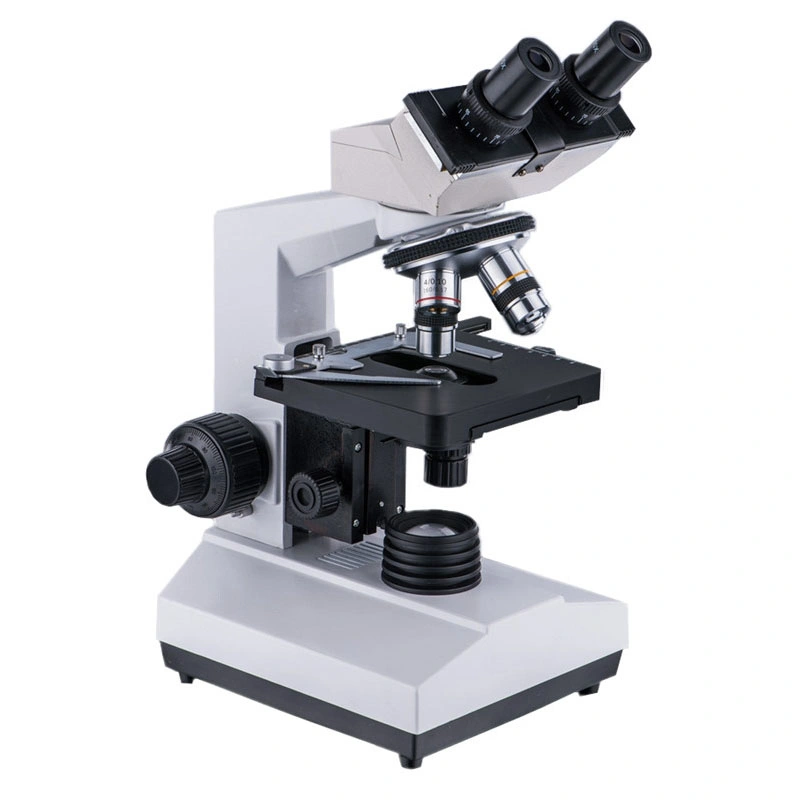
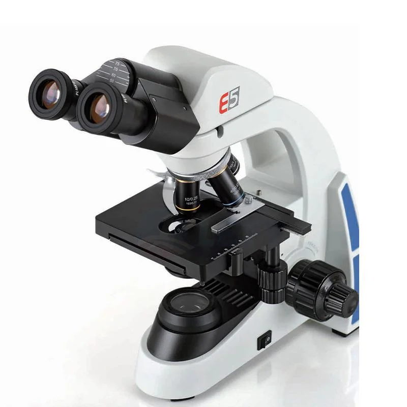
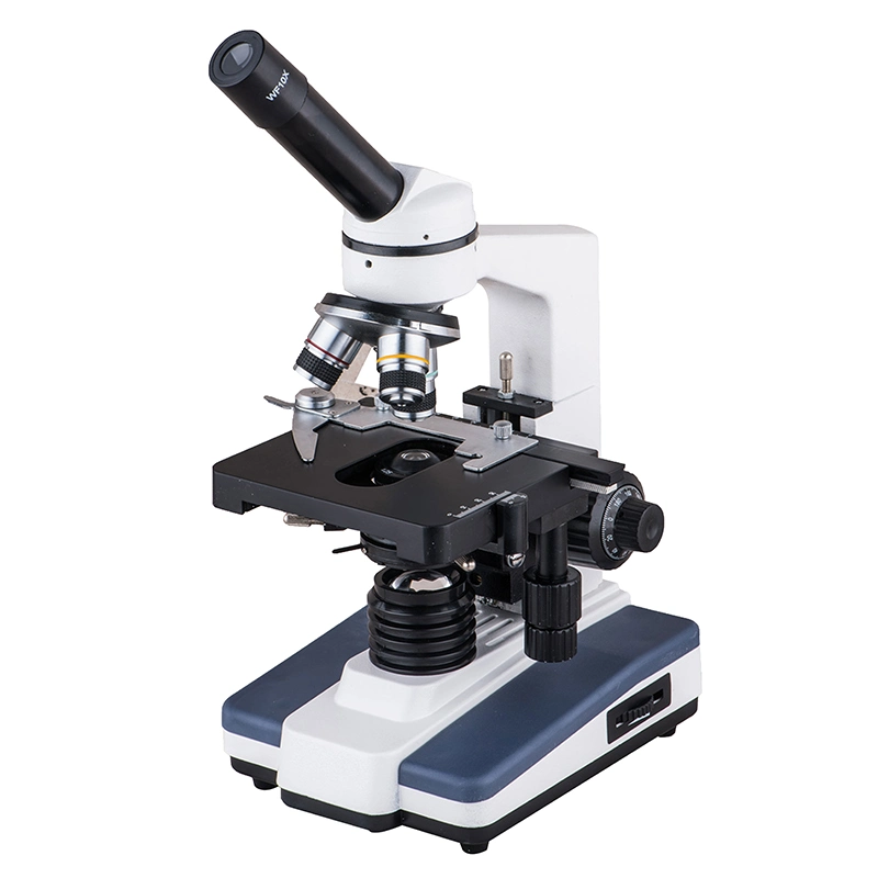
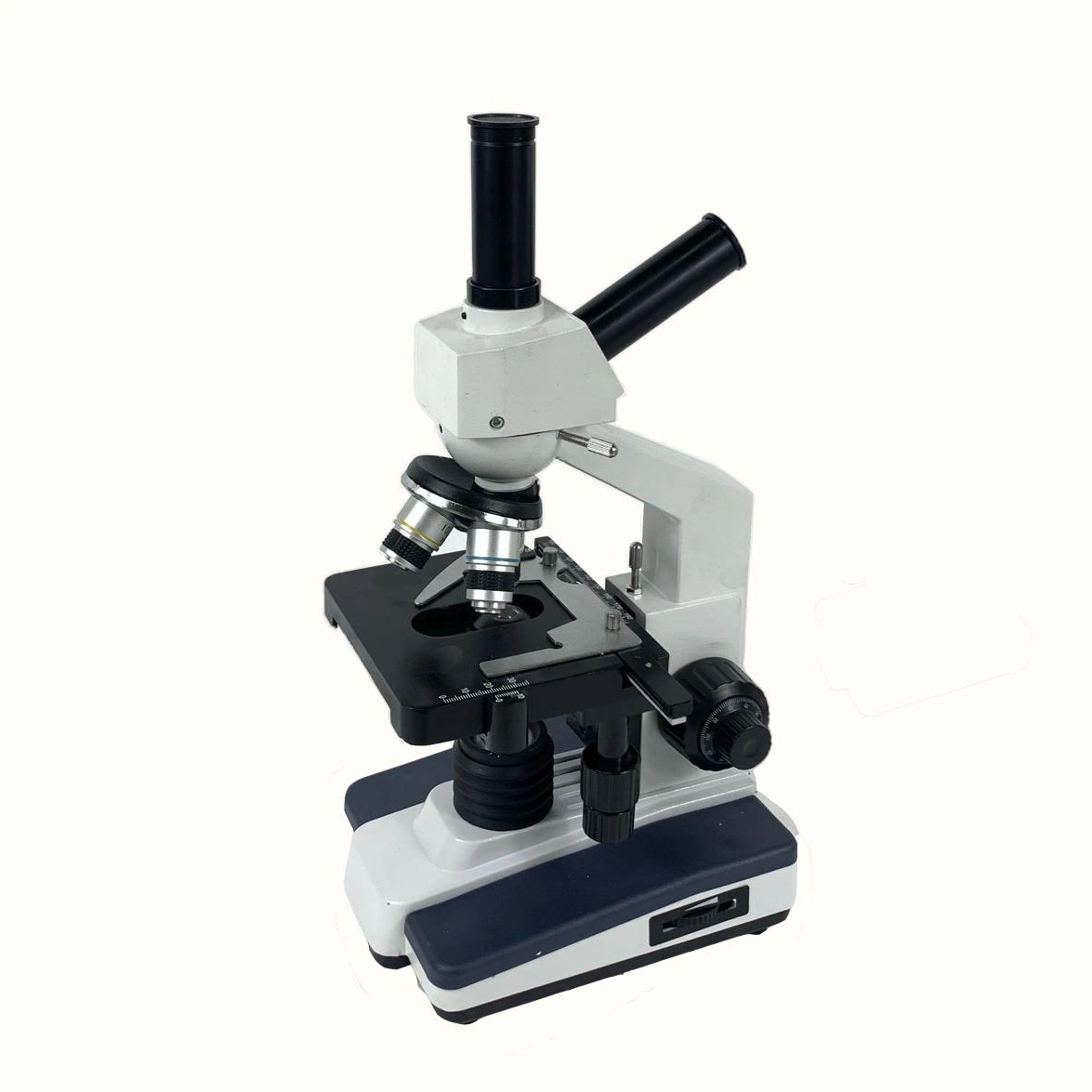
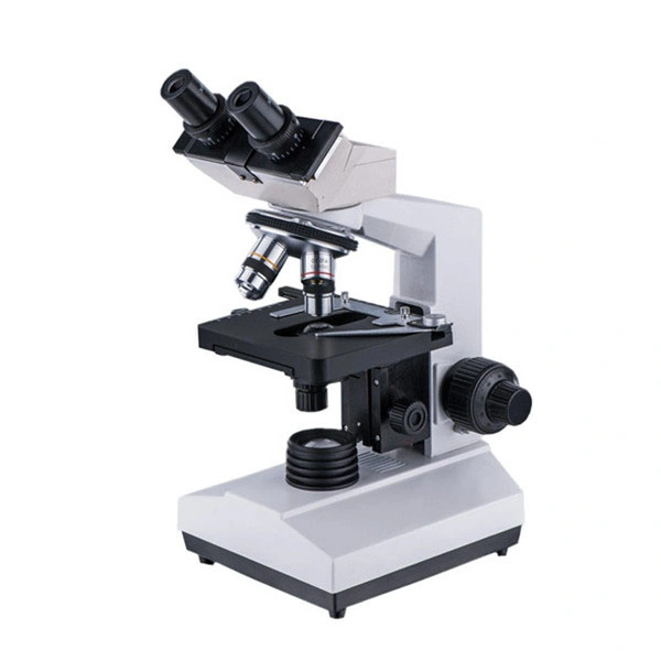
Medical Instrument Biological Stereo Microscope With CE XSP Series KS107V
IntroductionThis instruction manual is for the Biological Microscope XSP series.To ensurs the safety and obtain optimum performance and to familiarize yourself fully with use of this microscope,we recommend that you study this manual thoroughly before operating the microscope.Retain this instruction manual in an easily accessible place near the work desk for future reference.Operation Instruction- Insert the eyepiece into the eyepiece tube, and screw objectives into the nosepiece insequence of different magnification from low to high. Then put the specimen on the stage and secure it the position with tablet and move it to the center of stage.
- Turn on the power switch and adjust the brightness form dark to bright slowly. After working, you must adjust the brightness to a little dark before you turn it off.
- Observe the specimen from lower magnification objective firstly and move the specimen to the center of view field,then rotate higher magnification objective. You may use the fine focusing knob to obtain the clear image.When100X(s.oil)objective is used,you should fill up with cedarwood oil(without bubble)between the front of objective and the specimen surface. After working, it should be wiped with a few xylene immediately.
- In order to obtain bright and clear image, the illumination must be adjusted. When different objective is chosen,you should adjust the iris diaphragm of the condenser and different brightness of the light.
- When the lamp needs to replace,you should shut off the power switch and replace it after the lamp is cool. Note: The contact must be firmed,and the filament center should be adjusted.
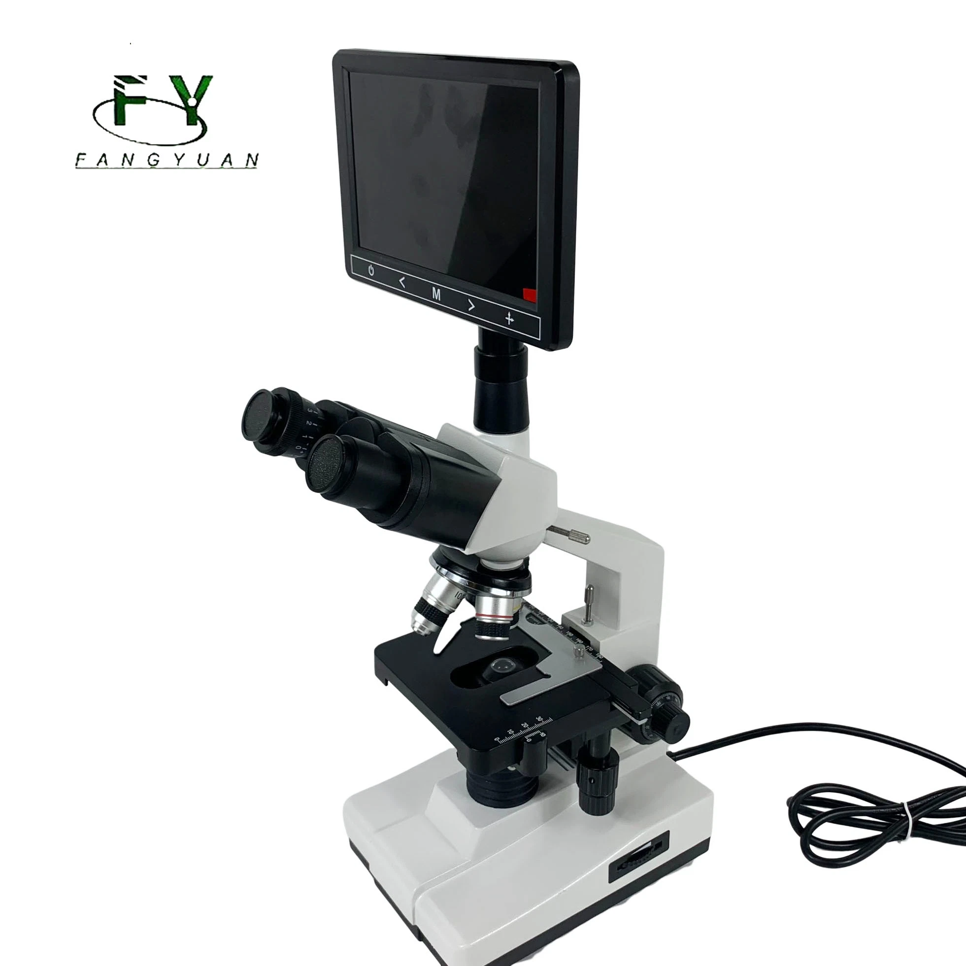
- If the camera or video attachment is used, you may insert the attachment into the tube of trinocular head of microscope,then adjustable tube until the image is clear enough.
SpecificationObjectives| Category | Magnification | Numerical aperture | Working distance | Remark |
Achromatic
objectives | 4X | 0.10 | 37.5 | |
| 10X | 0.25 | 7.613 | |
| 40X(s) | 0.65 | 0.632 | Spring |
| 100X(s.oil) | 1.25 | 0.198 | Spring |
Eyepieces| Category | Magnification | Diameter of view-field(mm) | Remark |
| Wide field eyepiece | WF10X | ф18 | |
| WF16X | ≥ф11 | |
Total magnification| Eyepiece/Total magnification/Objective | 4X | 10X | 40X(S) | 100X(S.oil) |
| 10X | 40X | 100X | 400X | 1000X |
| 16X | 64X | 160X | 640X | 1600X |
Mechanical tube length:160mm
Object to primary image distance:195mm
Stage size:140X140mm,moving range:75X45mm
ABBE condenser, NA=1.25 with iris diaphragm(Kohler illumination is optional)
Coarse/fine focal range:30mm
Illumination:6V 20W halogen lamp, adjustable brightness
Power: AC input 220V ±22V 50HZ±110V,Dc output 6VPhysical CharateristicsSize:185*270*400mmGross Weight:6.5KG Product show
About Us
Any details please contact us!
 Complaint