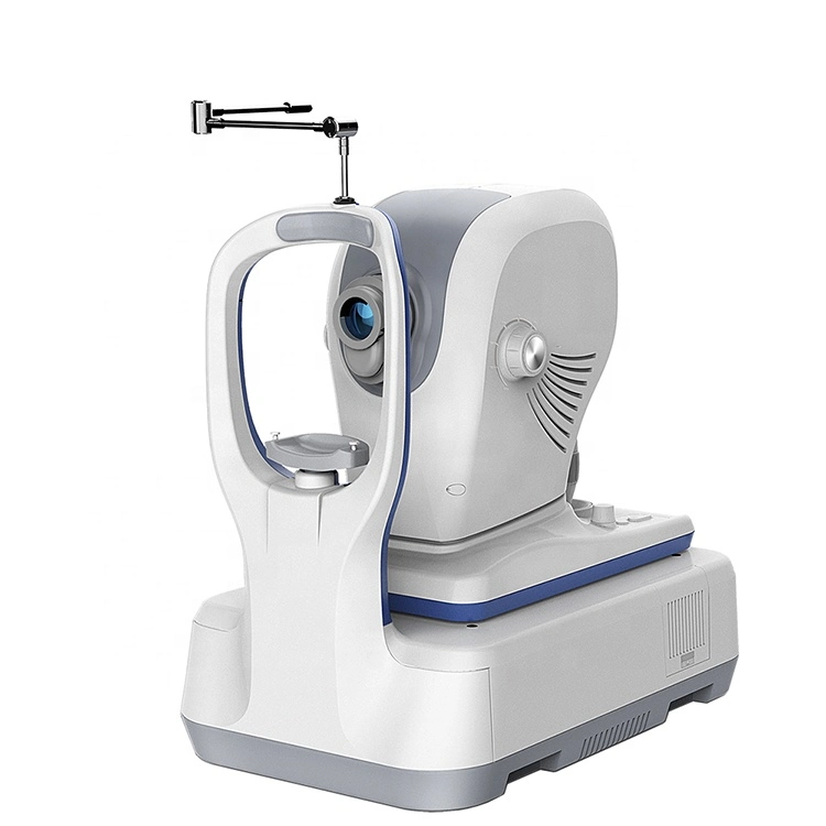
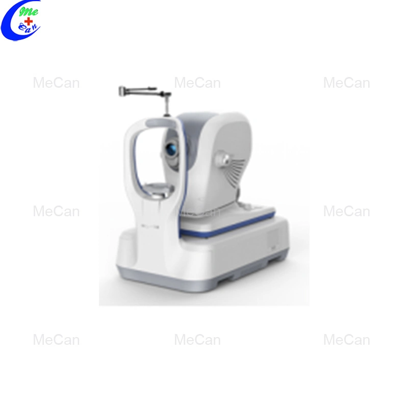
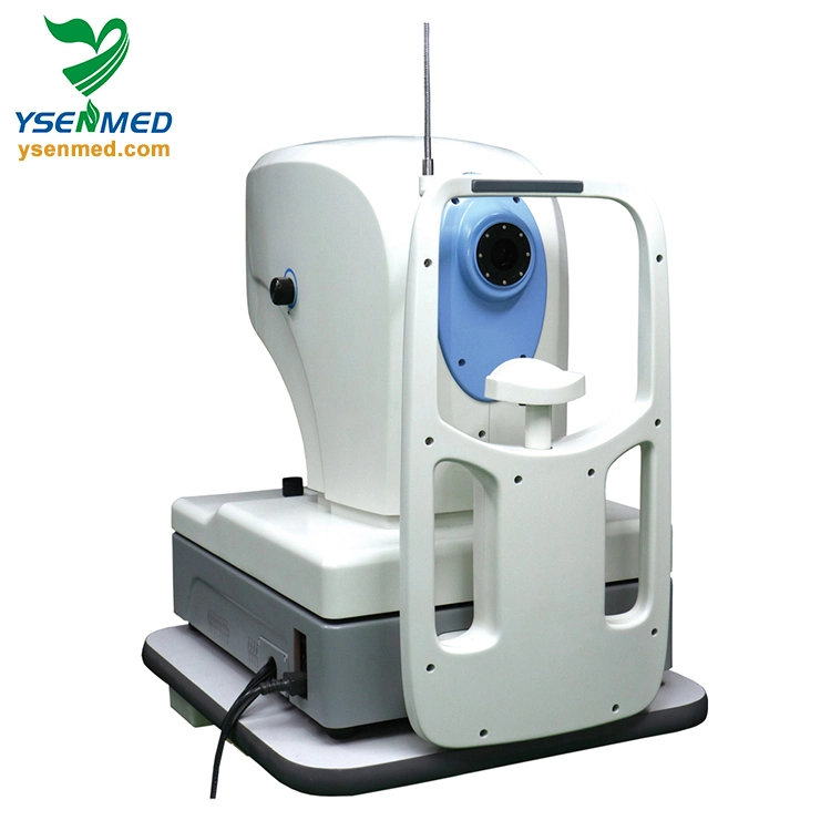
Ysoct500 Medical Ophthalmology Optical Coherence Tomography Scanner

Min.Order: 1
Product origin: Guangzhou, Guangdong, China
Infringement complaint: complaintComplaint
US$ -1

Description
YSOCT500 medical ophthalmology optical coherence tomography scanner


Technical Data

Feature
Remote data transmission system is professional software developed by us, aimed at digital storage and management for ophthalmic imaging examination. In the circumstances, YSOCT500 can implement functions of cloud storage of data and distributed resource sharing, so that doctors can read and analyze cases, and carry out telemedicine, etc..

Product Introduction
The first OCT with fundus color photography
YSOCT500 OCT with fundus color photography with variable retina scanning modes and analysis procedures, which provides direct and reliable analysis reports for auxiliary 
examinations of retina diseases and glaucoma.

High- definition and comprehensive 

High-definition OCT images with clear color fundus photography present a more accurate details in scanning area.
OCT scanning for macula
Fundus color photography


Fundus disease
Analysis for macula thickness

3D scanning in macula area of 6mm obtains more than 128 OCT images and mass data. And through professional calculated software, topographic map of retina thickness is got to analyze quality and quantity directly and precisely for diagnosis of retina diseases.

Analysis for macula lesion followup 
Macula lesion follow-up visit analysis software helps doctors to judge progress of disease and effect of therapy, such as followup of age-related macula lesion, cystoid macular edema therapy comparison before and after, etc..


Glaucoma

Comprehensive analysis for optic disc and nerve fibers of the retina

Scanned for optic disc and nerve fiber levels of retina (RNFL), it obtains RNFL images and related specifications of optic disc, such as rim area, cup to disc ratio, etc. and offers reliable quantitative analysis data for glaucoma diagnosis.

Quantitative analysis for optic disc: obtain cup to disc ratio and rim area by scanning for optic disc region

Quantitative analysis for RNFL thickness of two eyes: include TSNIT thickness profile images, RNFL quadrantal diagram and hour images. Compare with normal value database to judge changes of NFL thickness.


specification    YSOCT500
parametertechnologySpectral Domain OCT
light soruce830nm
 horizontal resolution15um
vertical resolution5um
scan deepth2.5mm
scan speed270000A-36000A
pupil size3mm
Scan method 3D scan ; Raster 2048x32;circle2048x32 ;radial 2048x32;HD scan;HD 5-line
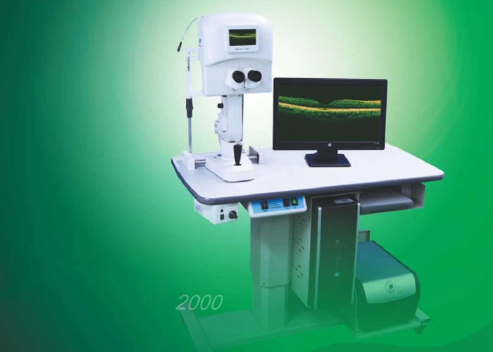
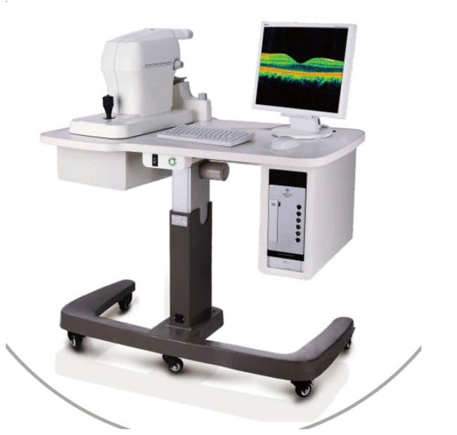
softwaremacular  Retina thickness analysis;3D view;Binoculus comparison, follow-up analysis
glaucoma  RNFL analysis; cup-disc analysis; progressive analysis;
anteriorconrneal map and thickness, chamber analysis
fundus  with fundus camera function


Product Tag:
Related categories:
Scroll to Top