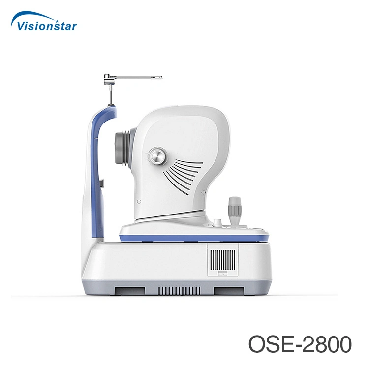
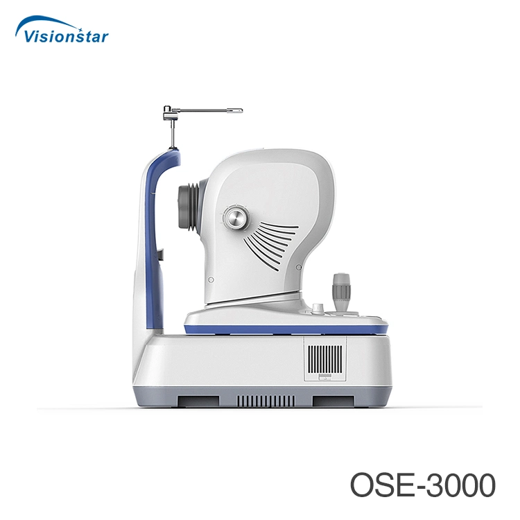
 Complaint

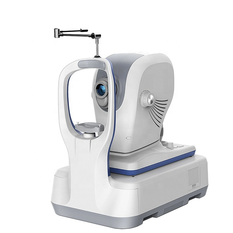
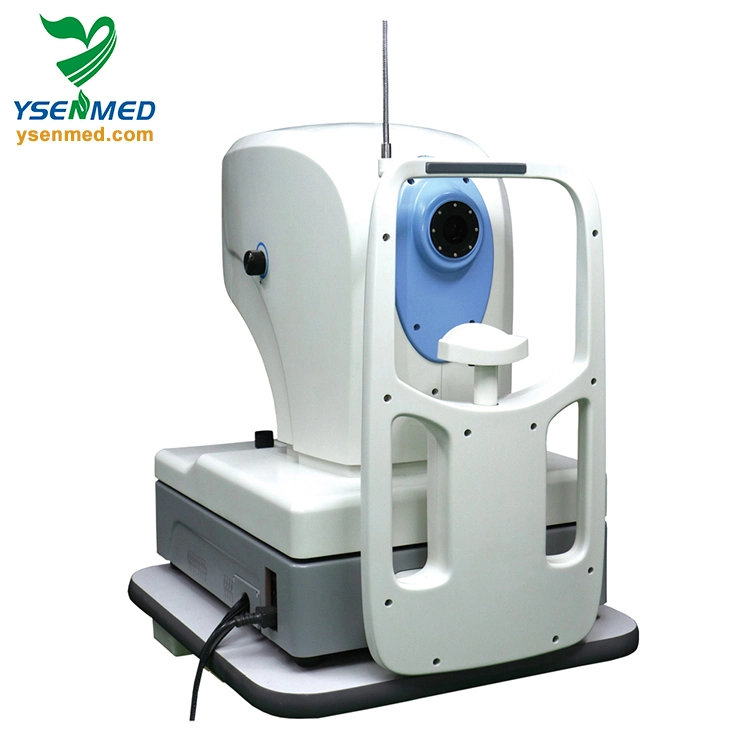
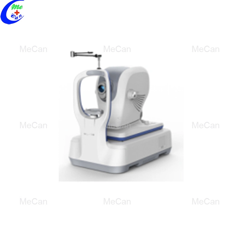
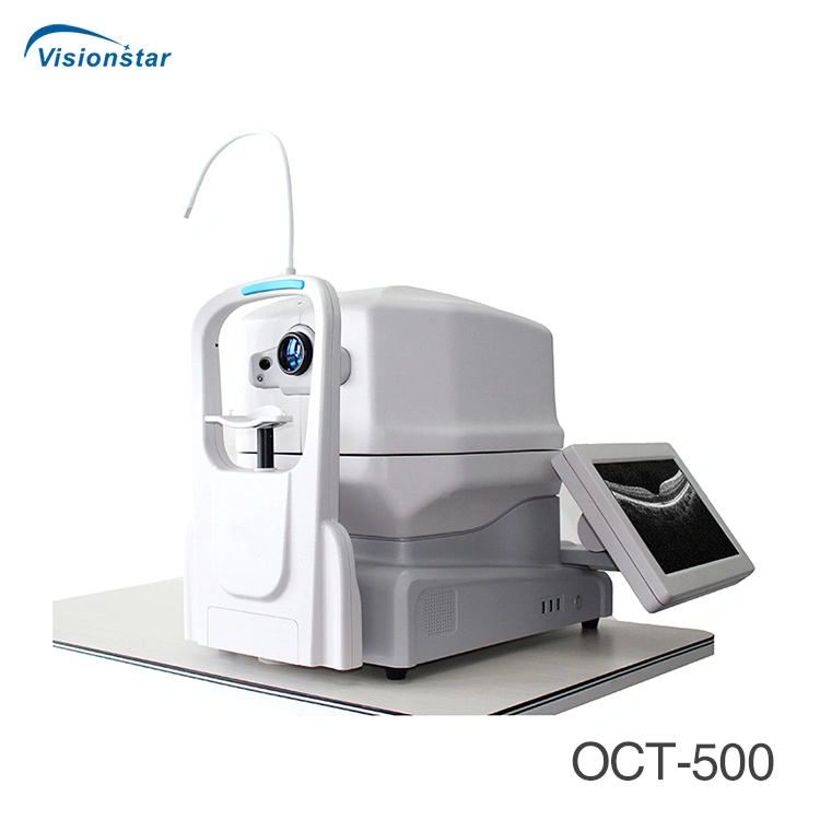
OCT IMAGING | ||
Methodology | Spectral domain OCT | |
Optical source | Super luminescent diode (SLD), 840 nm | |
Scan speed | 80,000 A-scans/s | |
Axial resolution (optical) | 5 microns (optical), 3.6 microns (digital) | |
Transverse resolution | 15 microns (optical), 3 microns (digital) | |
A-scan depth | 3 mm | |
Diopter range | - 20 to + 20 diopters | |
Scan patterns | Macular: HD line scan (6 / 12 mm), 3D scan (6 mm x 6 mm), 6-line radial scan, Multi (X-Y: 5 x 5) Disc: 3D scan (6 mm x 6 mm) Anterior: HD line scan (6 / 16mm), 6-line radial scan | |
FUNDUS IMAGING | ||
Methodology | Line scanning laser ophthalmoscopy (LSLO) | |
Minimum pupil diameter | 3.0 mm | |
Field of view | 45 degrees | |
VASCAN™ OCTA MODULE | ||
| VASCAN Advance | VASCAN Essential |
Scanning volume/area | 3mm x 3mm 256 x 256 A-scans 6mm x 6mm 360 x 360 A-scans 8mm x 8mm 360 x 360 A-scans 12mm x 8 mm 540 x 360 A-scans | 3mm x 3mm 256 x 256 A-scans 12mm x 8 mm 540 x 360 A-scans |
Algorithm | C-OMAG | C-OMAG |
Segmentation options | Encoded, Vitreousretina Intrerface(VRI), Superfcial retina, Deepfcial retinal, Avascular, Choriocapillaris, Choriod, Custom | |
Quantitative analysis | Yes | Not available |
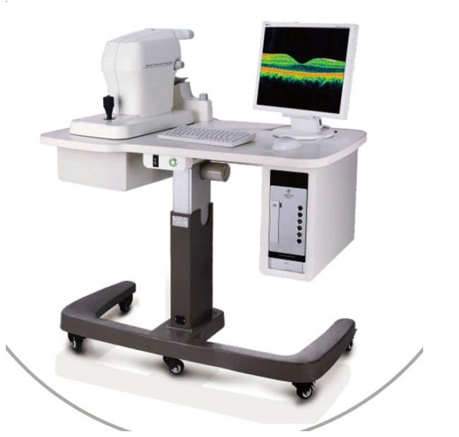
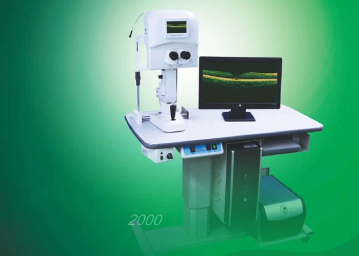
ELECTRICAL AND PHYSICAL | ||
Weight | 30.5 kg | |
Dimension | 532 mm (L) x 360 mm (W) x 540 mm (H) | |
Source voltage | AC 100 - 240 V, 50 Hz - 60 Hz | |
Power input | 90VA | |