 Complaint

| Option 1:2-5Mhz Convex probe |
| Option 2:5-8.5MHz Trans-vaginal probe |
| Option 3:5.5-9MHz Linear probe |
| Option 4:7.5 MHz rectal Linear Probe |
| Option 5:4-7Mhz micro convex probe |
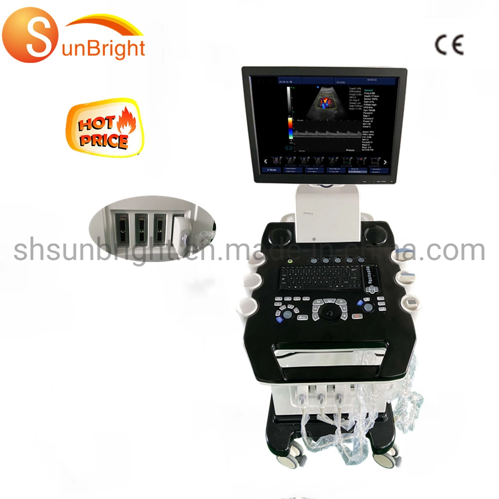
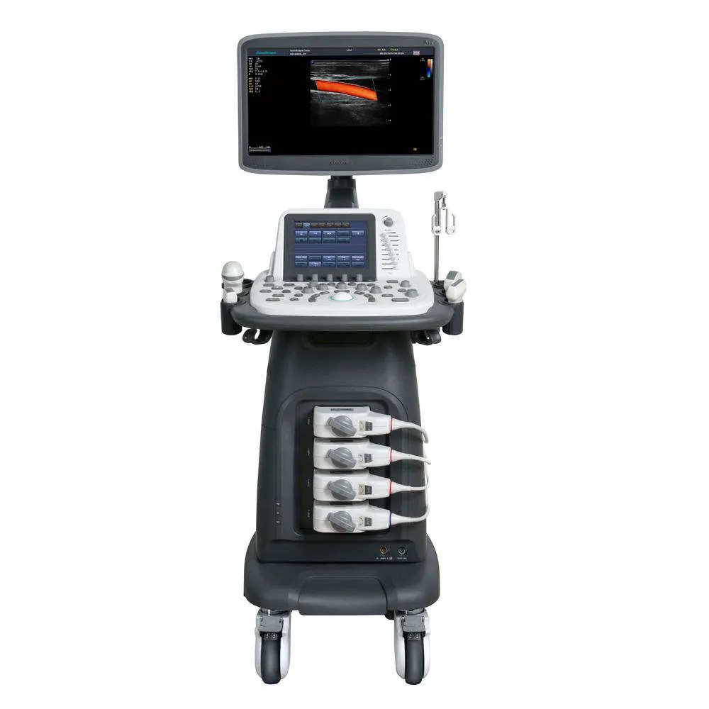
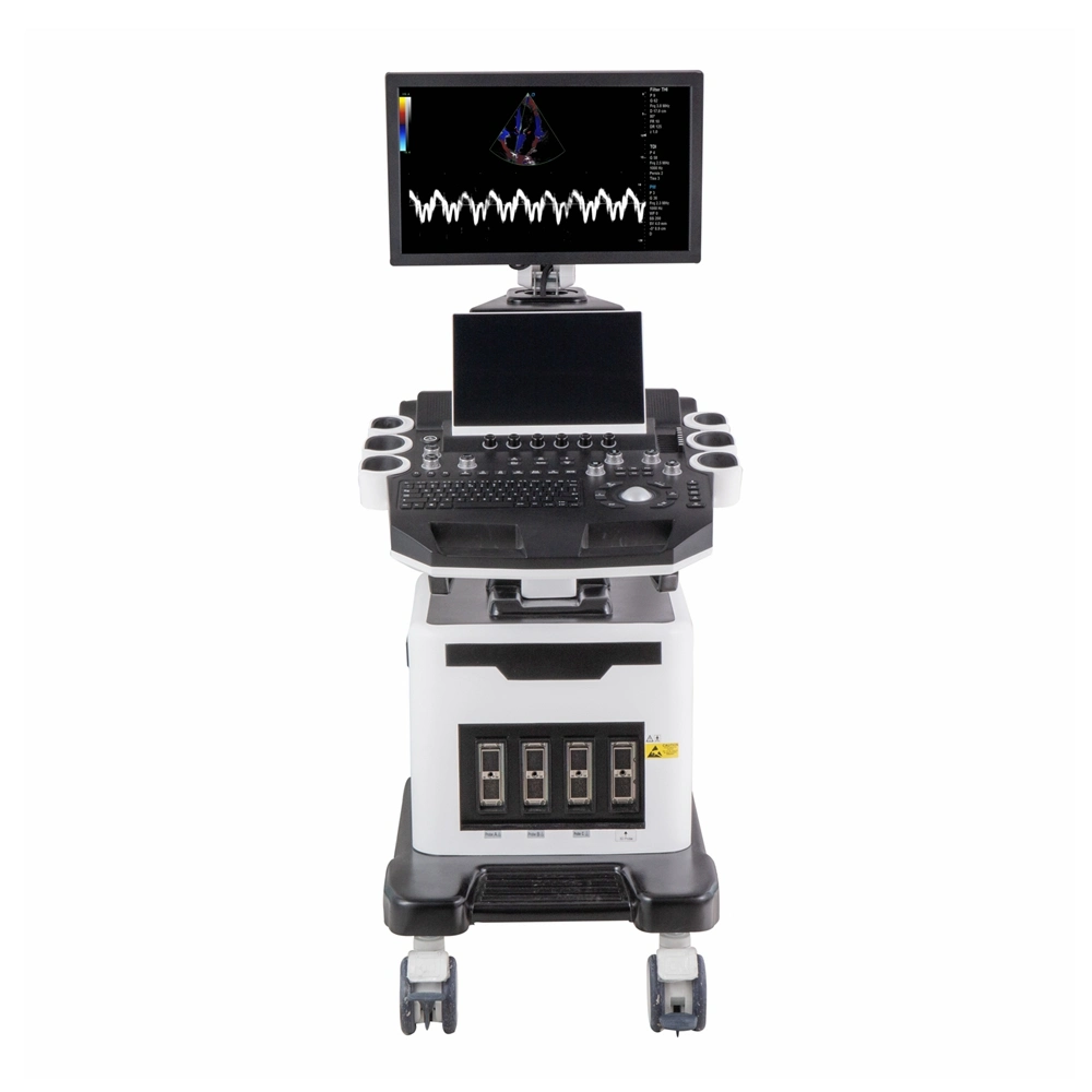
| Option 6: Trolley |
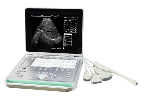
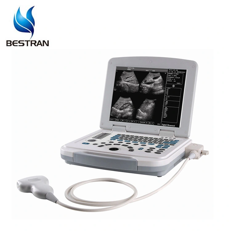
| 1, | Description of equipment: mainly used in abdomen, Urology, obstetrics and Gynecology, pediatrics, etc. |
| 2, | Functional characteristics |
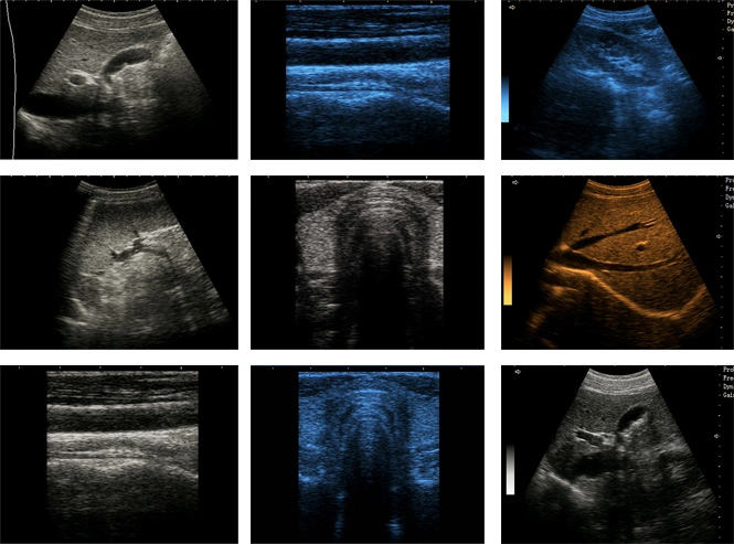
| 2.1 | All digital ultrasonic diagnostic instrument with high definition and rich function |
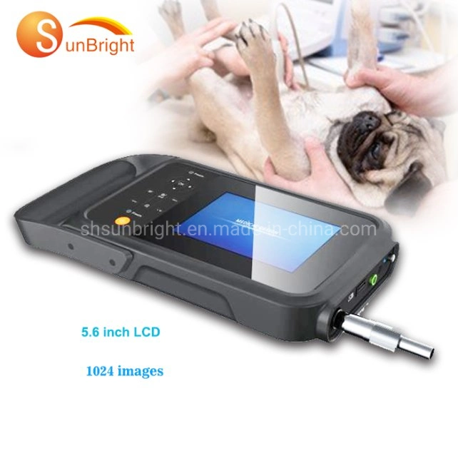
| 2.2 | The image is clear, easy to operate, and strong in endurance. It has great superiority in consultation in cities, townships and outdoor environments. |
| 2.3 | A variety of charging methods ensure more consultation in different environments. |
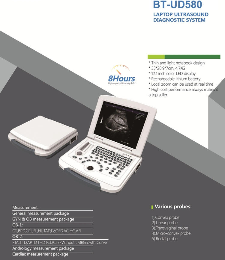
| 2.4 | 3-4 hours extra long standby time |
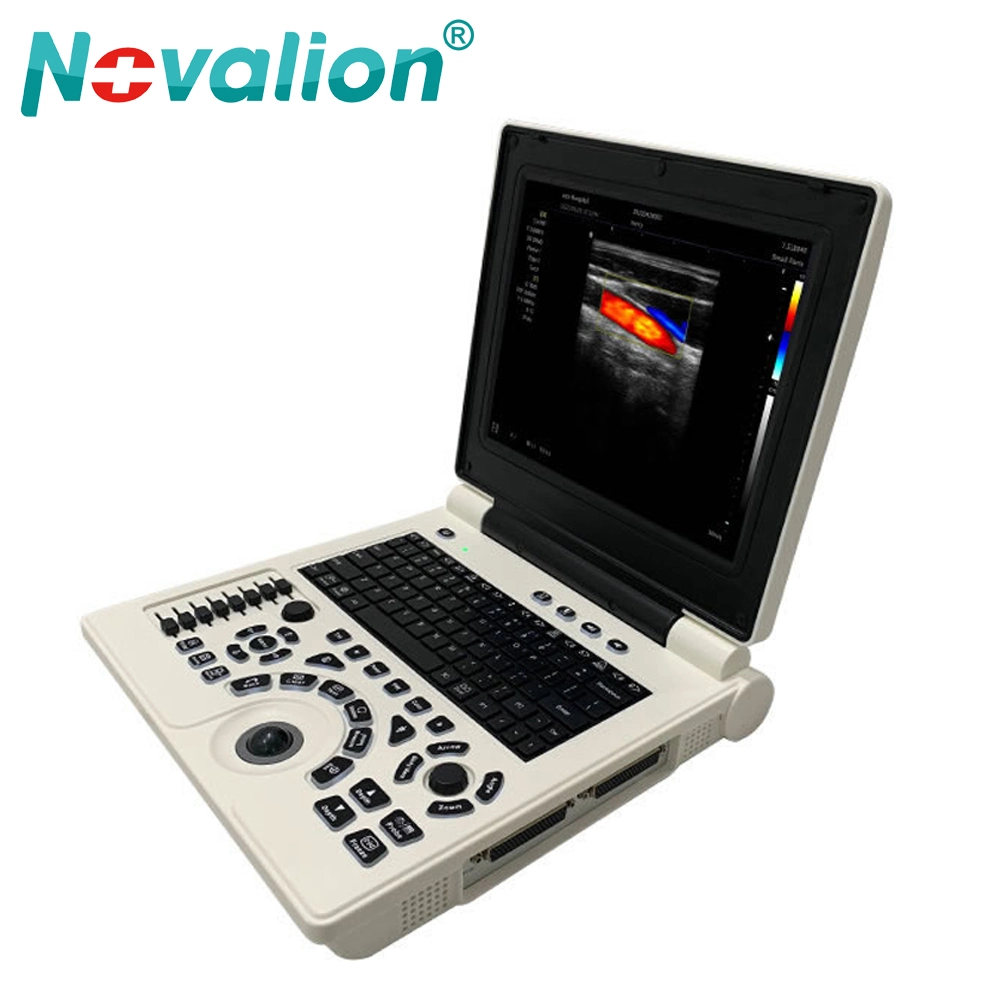
| 2.5 | 12.1 inch LED liquid crystal display |
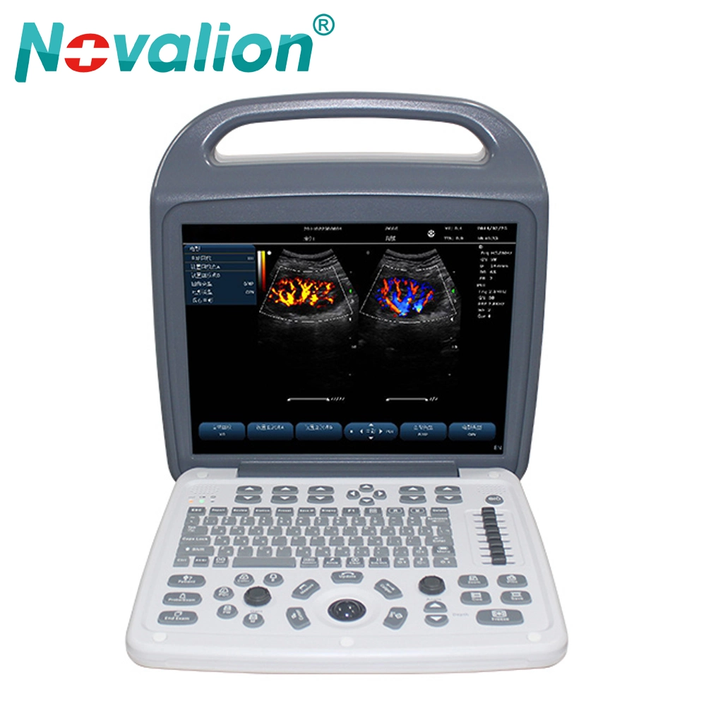
| 2.6 | Powerful image post-processing functions |
| 2.7 | The first class digital imaging technology, the image is clearer |
| 2.8 | DBF all digital beamforming |
| 2.9 | DRF real-time dynamic reception of focus by point by point |
| 2.10 | DRA real-time dynamic sound velocity change |
| 2.11 | THI tissue harmonic imaging |
| 2.12 | RDA real-time dynamic aperture imaging |
| 2.13 | DFS numerical control dynamic frequency scanning |
| 2.14 | RDF real-time dynamic filtering |
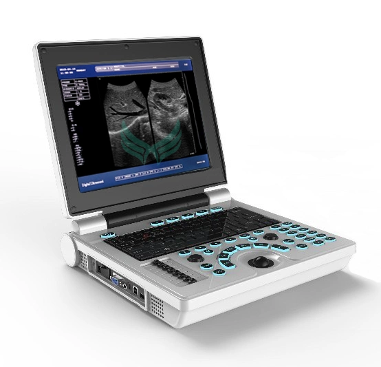
| 2.15 | A stable and concise operating platform |
| 2.16 | Backlight silicone keyboard, more comfortable and wearable, darkroom use no longer worry |
| 2.17 | Intelligent menu, human-computer dialogue is easy and quick |
| 2.18 | Shows two puncture guide lines, adjustable angles and positions. |
| 2.19 | The multiple rate shows that the diagnosis is more accurate. |
| 2.20 | External USB storage, image uploading more convenient |
| 2.21 | Large volume movie playback, image automatic circulation demonstration |
| 2.22 | Abundant measurement functions: distance, circumference, area, volume, obstetric table, heart software package, etc. |
| 3, | Performance introduction |
| 3.1 | Display: 12.1 inch LED medical display |
| 3.2 | Scanning mode: convex matrix / linear array / microconvex |
| 3.3 | Probe interface: Standard 1, 2 (optional), automatic identification function, support multiple probe work. |
| 3.4 | Interface: Chinese / English interface |
| 3.5 | Display mode: B, B+B, 4B, B+M, M |
| 3.6 | Electron focusing: four segment electron focusing |
| 3.7 | Postural markers: 97 species |
| 3.8 | 3.5MHz: 2.0MHz,2.5MHz,3.5MHz,4.0MHz,5.0MHz 5.0MHz: 4.0MHz,4.5MHz,5.0MHz,6.5MHz,7.0MHz 6.5MHz: 5.0MHz,5.5MHz,6.5MHz,7.5MHz,8.5MHz 7.5MHz: 5.5MHz,6.5MHz,7.0MHz,7.5MHz,9.0MHz |
| 3.9 | Image mirroring: upper and lower mirrors, left and right mirrors, black and white flip, in any mode can be mirrored conversion, operation |
| 3.10 | The image can be rotated at 0 degrees, 90 degrees, 180 degrees, 270 degrees, and 360 degrees. |
| 3.11 | Measurements: General measurement, GYN & OB measurement package, Andrology measurement , Cardiac measurement. distance, circumference, area, volume, heart rate, OB-1:GS,BPD,CRL,FL,HL,TAD,LV,OFD,AC,HC,AFI OB-2: FTA,TTD,APTD,THD,TCD,CI,EFW,Input LMP,Growth Curve |
| 3.12 | The function software package and measurement method can be selected through the track ball cursor. |
| 3.13 | Angle measurement, you can intuitively see the angle and length of the angle. |
| 3.14 | Have histogram function |
| 3.15 | Have depth measurement function |
| 3.16 | It has 16 obstetric measurement packages and has fetal growth curve function. |
| 3.17 | In any imaging mode, the data can be measured in real time |
| 3.18 | Automatic memory and generative function of obstetric data |
| 3.19 | Character display: date, clock, name, gender, age, doctor, hospital, annotation (full screen character editing) |
| 3.20 | One key out of patient information entry function |
| 3.21 | Movie playback: 512 frames, continuous playback or one by one view. |
| 3.22 | Permanent storage: built in 8G memory, more than 4000, supporting U disk storage (available for storage). |
| 3.23 | Single memory image time is less than 6 seconds |
| 3.24 | A key to find saved images, can be found by the date named folder quickly found and a key export. |
| 3.25 | Gray scale: 256 level |
| 3.26 | With the function of puncture and guidance, the angle and position of the two puncture lines can be adjusted visually. |
| 3.27 | Lithotripsy positioning, dynamic target tracking function |
| 3.28 | Dynamic range: 0-135dB, step 8, with independent buttons, visually adjustable, adjustable cycle. |
| 3.29 | Intelligent TGC control: 8 segments. |
| 3.30 | Total gain: 0-100, step 2 |
| 3.31 | Variable aperture, dynamic trace, dynamic digital filtering, etc. |
| 3.32 | 2 stage visible tunable harmonics of tissue |
| 3.33 | 8 kinds of fake color processing, etc. |
| 3.34 | The 4 frames are independent keys, visually adjustable, cycle adjustable, and can also be changed through the track ball cursor. |
| 3.35 | 6 kinds of line correlation, with independent buttons, visually adjustable, circular adjustment, or track ball cursor changes. |
| 3.36 | The 8 types of gamma correction are independent keys, visually adjustable, cycle adjustable, and can also be changed through the trackball cursor. |
| 3.37 | Edge enhancement 4 level adjustable, with independent buttons, visually adjustable, adjustable cycle, also can be changed through the track ball cursor. |
| 3.38 | The scanning range can be adjusted 4 levels, with independent buttons, visually adjustable, circular adjustment, and also can be changed by the track ball cursor. |
| 3.39 | Luminance key addition and subtraction enhancement and weakening function |
| 3.40 | Caps and Shift locking functions |
| 3.41 | Blind area: less than 4 |
| 3.42 | Maximum display depth:3.5MHz:307mm 6.5MHz:189mm 7.5MHz:166mm 5.0MHz:205mm |
| 3.43 | Geometric accuracy: transverse less than 5%, longitudinal less than 5% |
| 3.44 | Resolution: the side is less than 2mm, and the axis is less than 1mm |
| 3.45 | Interface: RS-232 interface, VGA interface, VIDEO interface, USB interface 2, DICOM |
| 3.46 | Display rate: 6 display modes; lesion diagnosis is more accurate. When it is above 1.5 and over 1.8, the depth can be enhanced. |
| 3.47 | Starting time is less than 10 seconds |
| 3.48 | Built-in 8800 Ma lithium battery |
| 3.49 | Power indicator light and charging indicator |
| 3.50 | Net weight 4.7KG |
| 3.51 | The button has a buzz sound, but open / close |
| 3.52 | Formulae of fast data measurement |
| 3.53 | You can quickly set time, name and other editors through the track ball and keyboard. |
| 4, | Standard: 12.1 inch LCD; 3.5MHz electronic convex array probe. |
| 5, | Accessories: 7.5MHz high frequency probe; 6.5MHz cavity probe; probe interface development dock; ultrasonic imaging workstation; portable trolley. |