Description
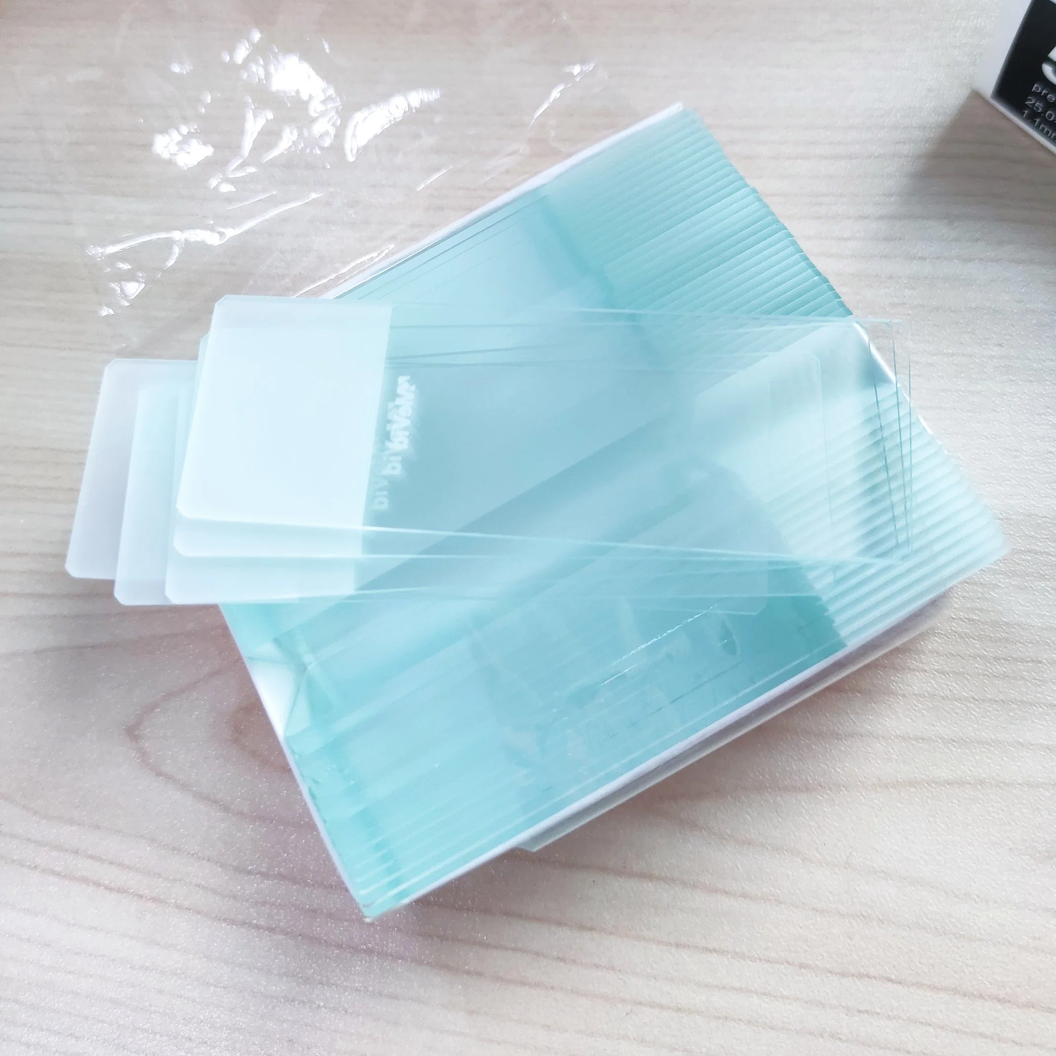
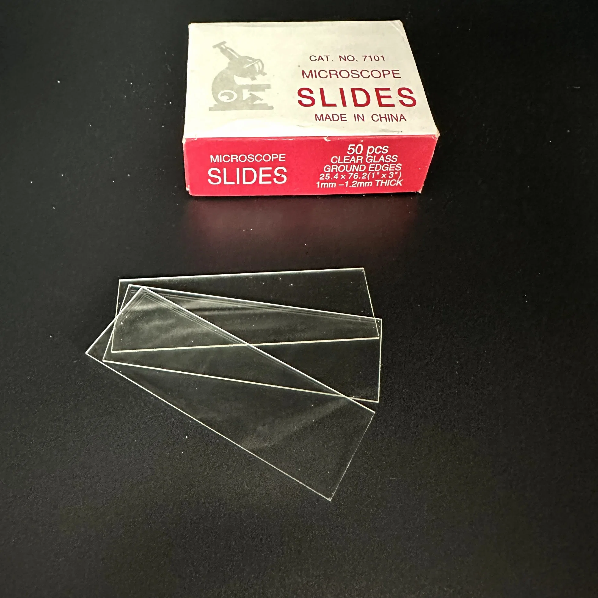
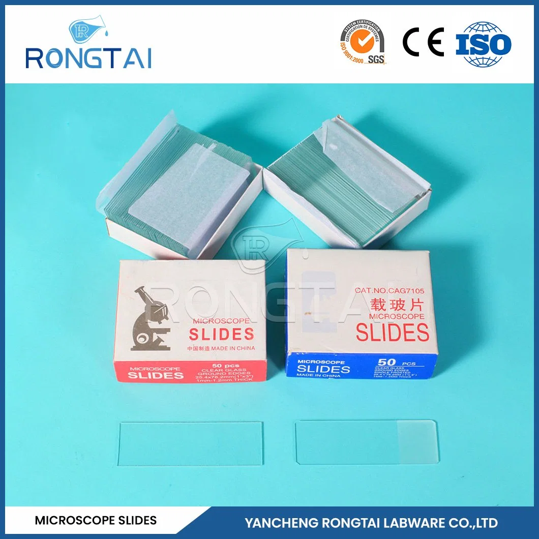
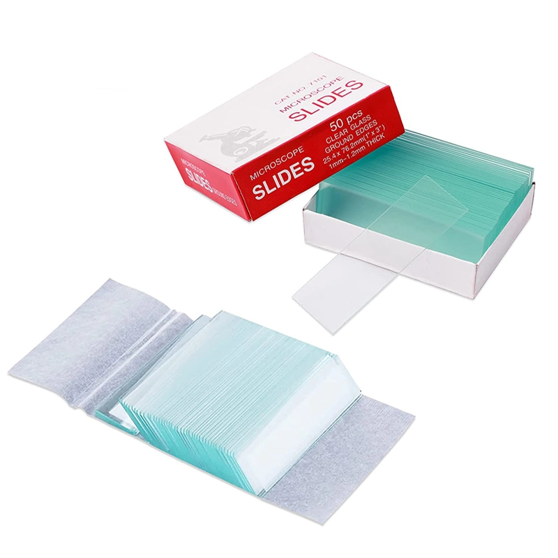
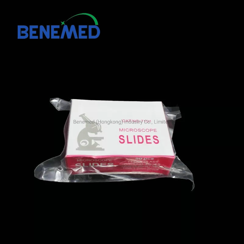
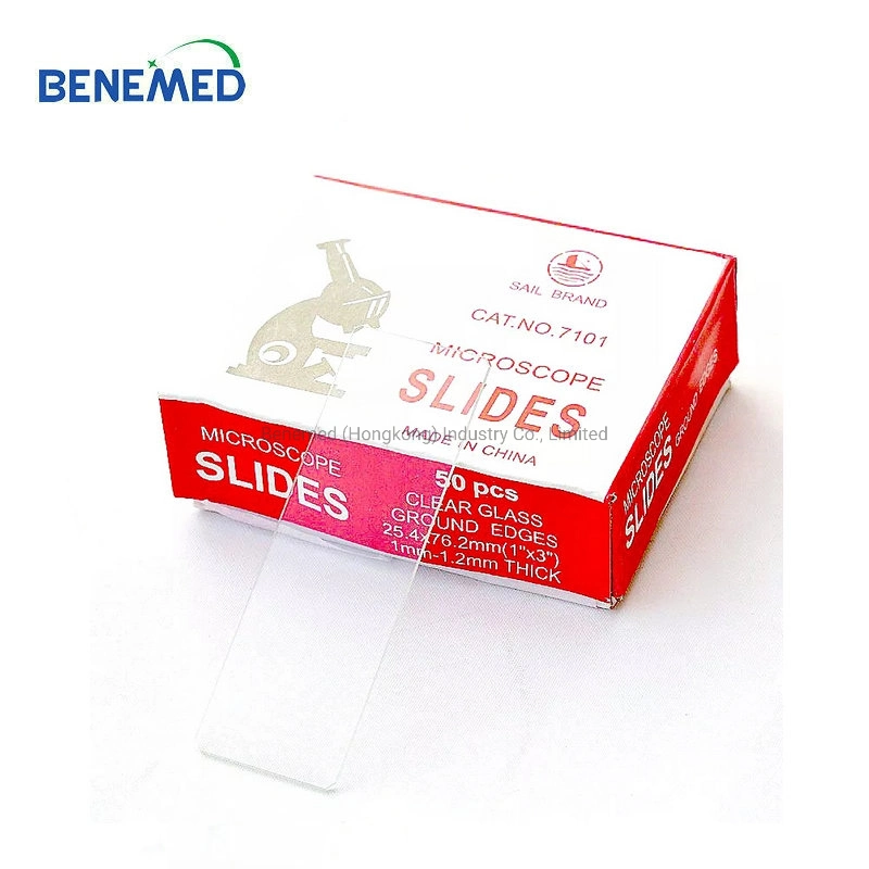
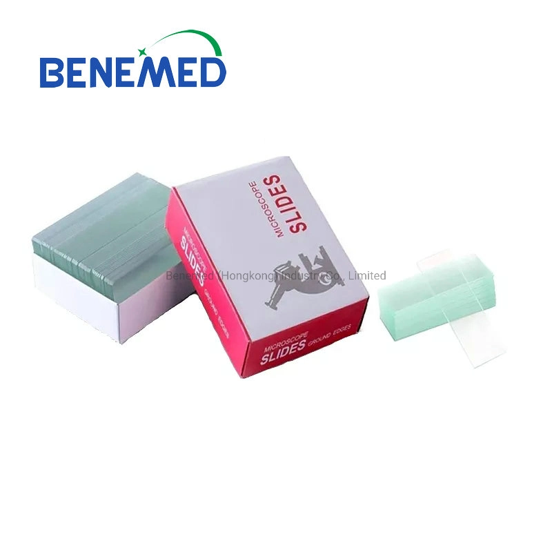
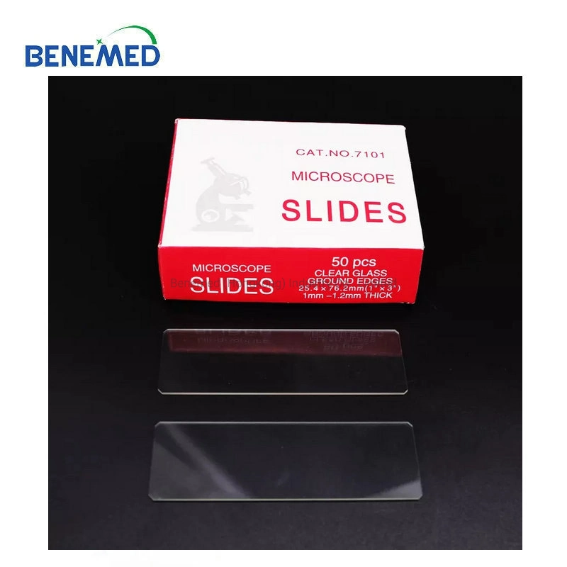
Laboratory Glassware Fine Quality Double Frosted Microscope Slides
| Glass microscope slide |
| Code | Size(mm) | Style | Material | Ctn size(cm) | Packing | GW(kg) |
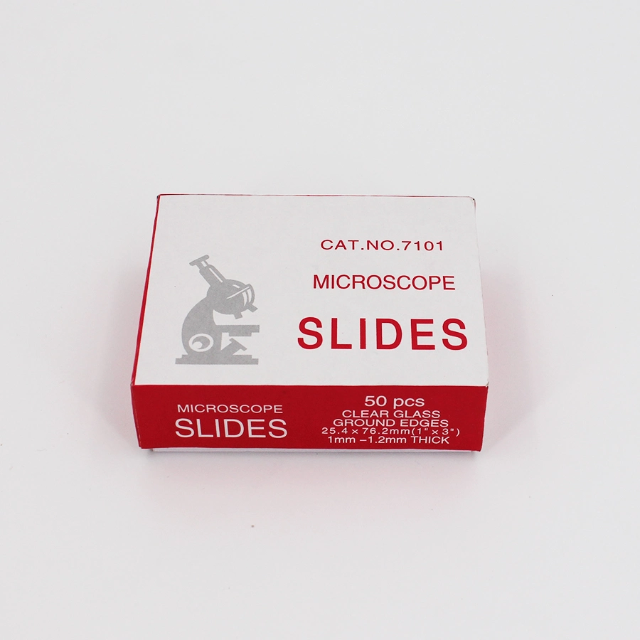
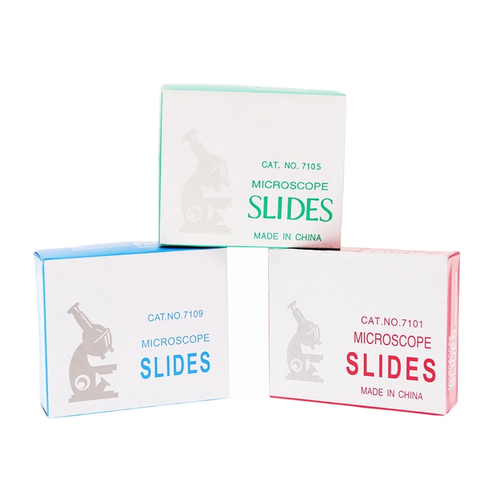
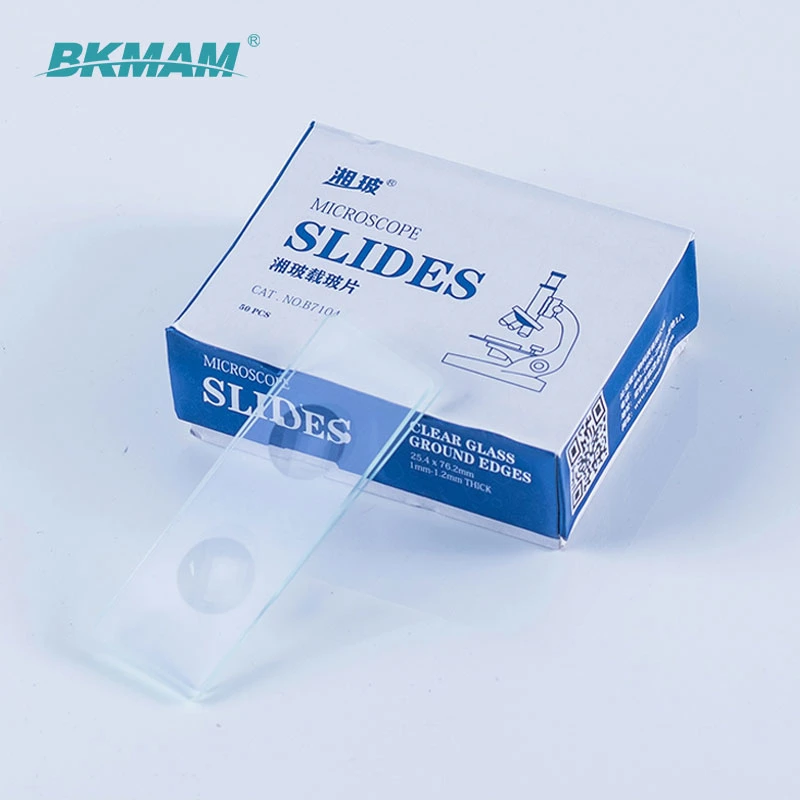
| TYI-7101 |
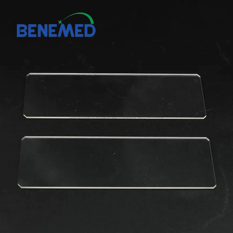
25.4*76.2 | Polished edge |
Glass |
37*18*16 |
50pcs.65pcs.72pcs/box,
50box/ctn |
13 |
| TYI-7102 | Cut edge |
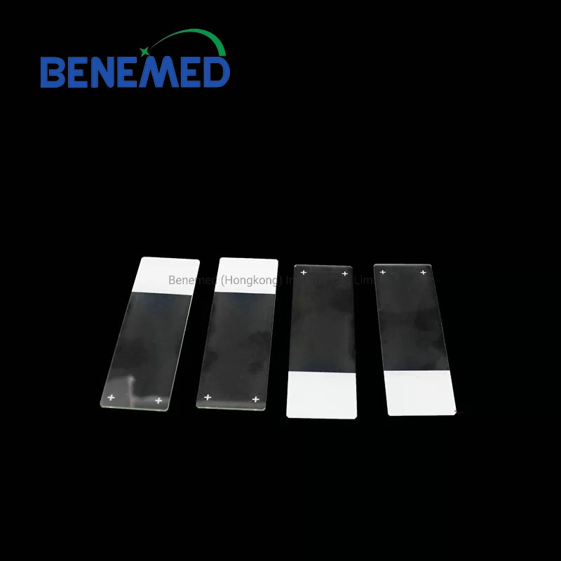
| TYI-7105 | Frosted on one slide |
| TYI-7107 | Frosted on double side |
Usage of the glass slide - The smear method is a method of filming the material evenly on the glass slide. Smear materials include single-celled organisms, small algae, blood, bacterial culture fluid, loose tissues of animals and plants, testis, anthers, etc. When smearing, pay attention to: (1) The glass slide must be clean. (2) The slides should be flat. (3) The coating must be uniform. Apply the droplet to the right of the middle of the slide and spread it evenly with a scalpel blade or toothpick. (4) The coating should be thin. Use another slide as a pusher, and gently push it from right to left along the surface of the slide with the smear drop (the angle between the two slides should be 30°-45°) to apply a thin layer evenly. (5) Fixed. For fixation, chemical fixatives or drying methods (bacteria) can be used for fixation. (6) Dyeing. Methylene blue is used for bacteria, Wright's stain for blood, and sometimes iodine. The dye solution should cover all the coated surfaces. (7) Rinse. Blot dry with absorbent paper or bake dry. (8) Coverslip. For long-term storage, cover with Canadian gum.
- The tablet pressing method is a tableting method in which the biological material is placed between the glass slide and the cover slip, and a certain pressure is applied to scatter the tissue cells.
- Mounting method is a method of sealing biological materials as a whole to make slide specimens. This method can be used to make temporary or permanent mounting. Mounting materials include: microscopic organisms such as Chlamydomonas, Spirogyra, amoeba, and nematodes; hydra, leaf epidermis of plants; wings, feet, mouthparts of insects, human oral epithelial cells, etc.
Attention should be paid to: (1) When holding the slide, it should be kept flat or placed on the platform. When dripping water, the amount of water should be appropriate, so that it is just covered by the coverslip. (2) The material should be spread out with a dissecting needle or tweezers without overlapping and flattened on the same plane. (3) When placing the coverslip, slowly cover the water droplets from one side to prevent bubbles from appearing. (4) When staining, place a drop of staining drop on one side of the coverslip and suck it from the other side with absorbent paper to evenly stain the specimen under the coverslip. After staining, in the same way, drop a drop of water, suck out the staining solution, and observe it under a microscope.
- Slices are slide specimens made from thin slices cut from living organisms. Due to different requirements, a razor blade can be used for free-hand sectioning, or the tissue block can be embedded in paraffin or collodion or frozen at low temperature and sliced with a microtome. Cut into 5-10 micron thin slices for optical microscope observation. Ultrathin sections cut from tissue blocks embedded in epoxy resin or methacrylic acid, with a thickness of 20-50 nanometers, are specially designed for observation under an electron microscope. Sections such as root tips and stems used in general teaching are generally called paraffin sections.
Cleaning method
Washing with water or alcohol, and then wipe it with cotton or gauze (it is best to use lens tissue, avoid touching the slide with your fingers, so as not to leave fingerprints on it, which will affect the next observation and use)
 Complaint