| Main components | Technique parameter | |
| IMD X-ray tube | Model:YSX-C35D | Model:YSX-C50D | |
| Power:3.5kW | Power:5.0kW | |
| Double focus fixed anode focus:1.5mm / 0.6mm | Double focus rotational anode focus: 0.3mm / 0.6mm | |
| Anode Heat Capacity:40kHu X-ray tube sleeve heat capacity:667kHu | Anode Heat Capacity:200kHu X-ray tube sleeve heat capacity:800kHu | |
| High Voltage X-ray Generator | Working frequency:40kHz | |
| Image intensifier | 9"(including 3 fields of view 4.5"/ 6"/9") | |
| Tube voltage | Fluoroscopy Radiograph:40kV-110kV | Fluoroscopy Radiograph:40kV-120kV | |
| Tube current | Fluoroscopy: 0.5-4mA Enhanced Automatic Fluoroscopy: 1mA-8mA Pulse Fluoroscopy :2pps;8mA | |
| mAs | 1mA.s-250mA.S | |
| Fluoroscopy mode | Manual Fluoroscopy; Semi-Automatic Fluoroscopy;Automatic Fluoroscopy; Enhanced Automatic Fluoroscopy; Pulse Fluoroscopy | |
| Intelligent exposure control | No matter the object is in the center or on the fringing field of the image intensifier, the images are the same. The exposure dose is reduced. | |
| Iris beam limiting device | when not exposure, keep in the state of Iris Beam limiting preview ,when exposure,keep blanking cycle track | |
| |
| General radiography | 40kV-120kV;1.0mA.s-250mA.s | step length 1 kV;1.0mA.s | |
| Digital radiography (digital spot film) | 40kV-120kV;Max 16mA | Common screen film radiography is eliminated. Digital radiography can get better images | |
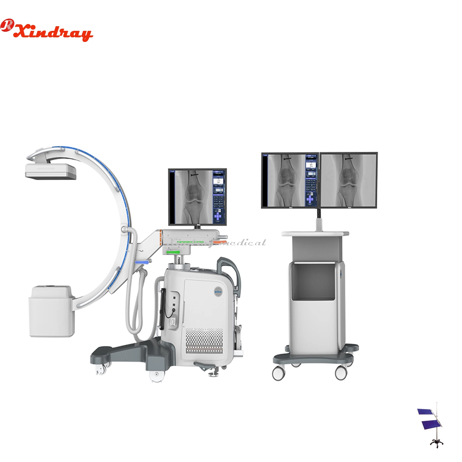
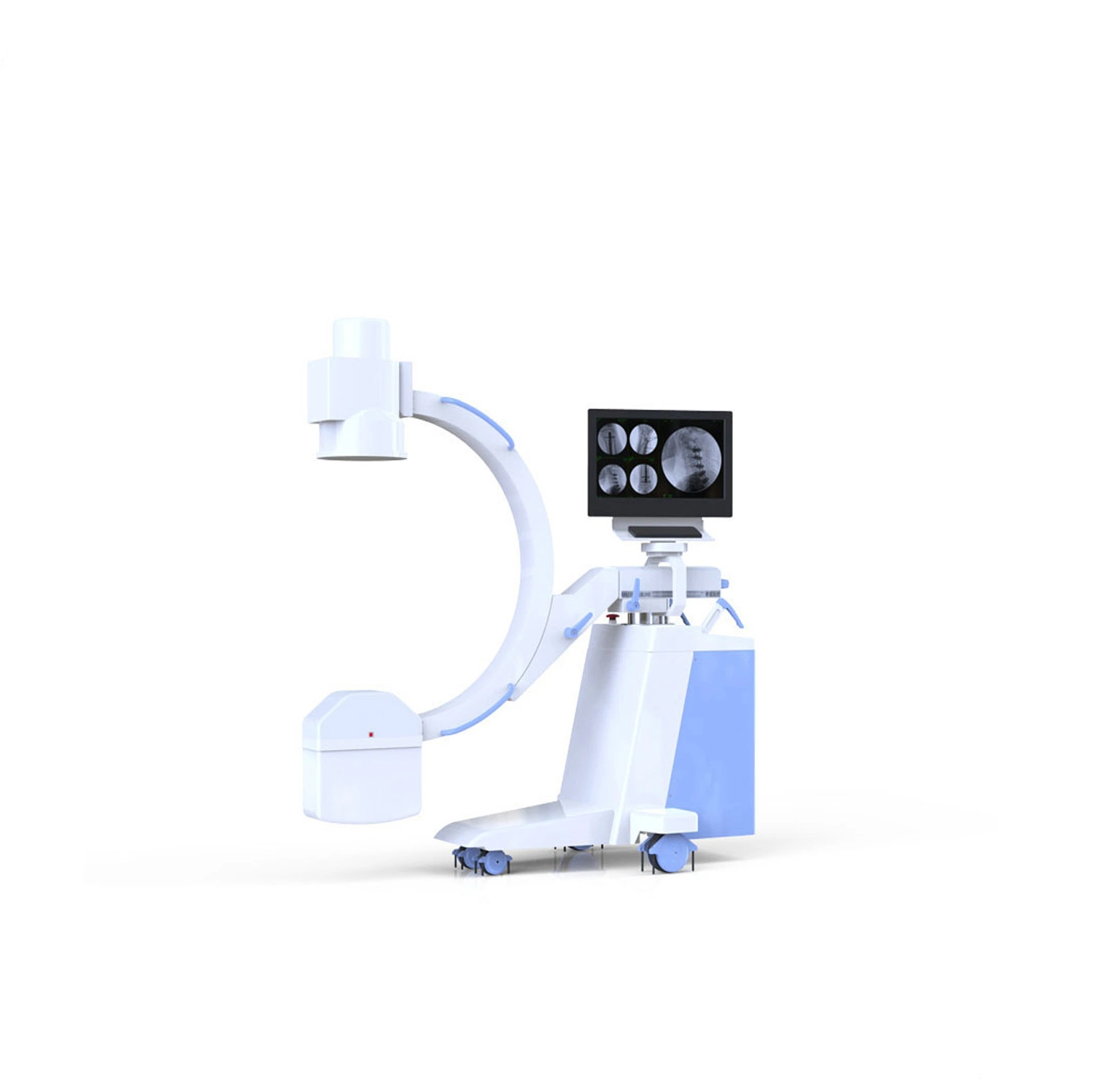
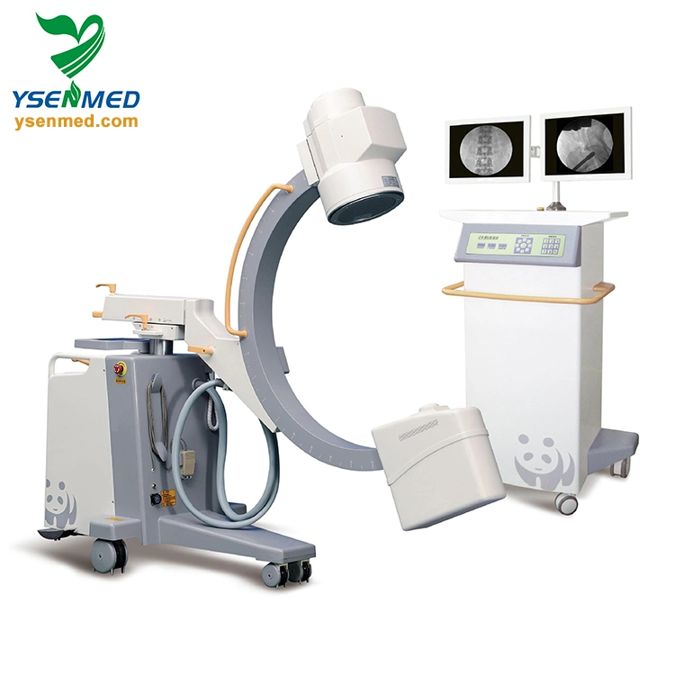
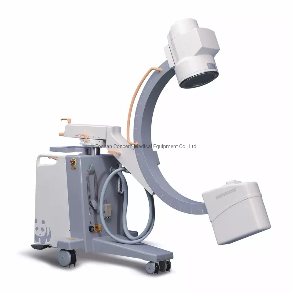
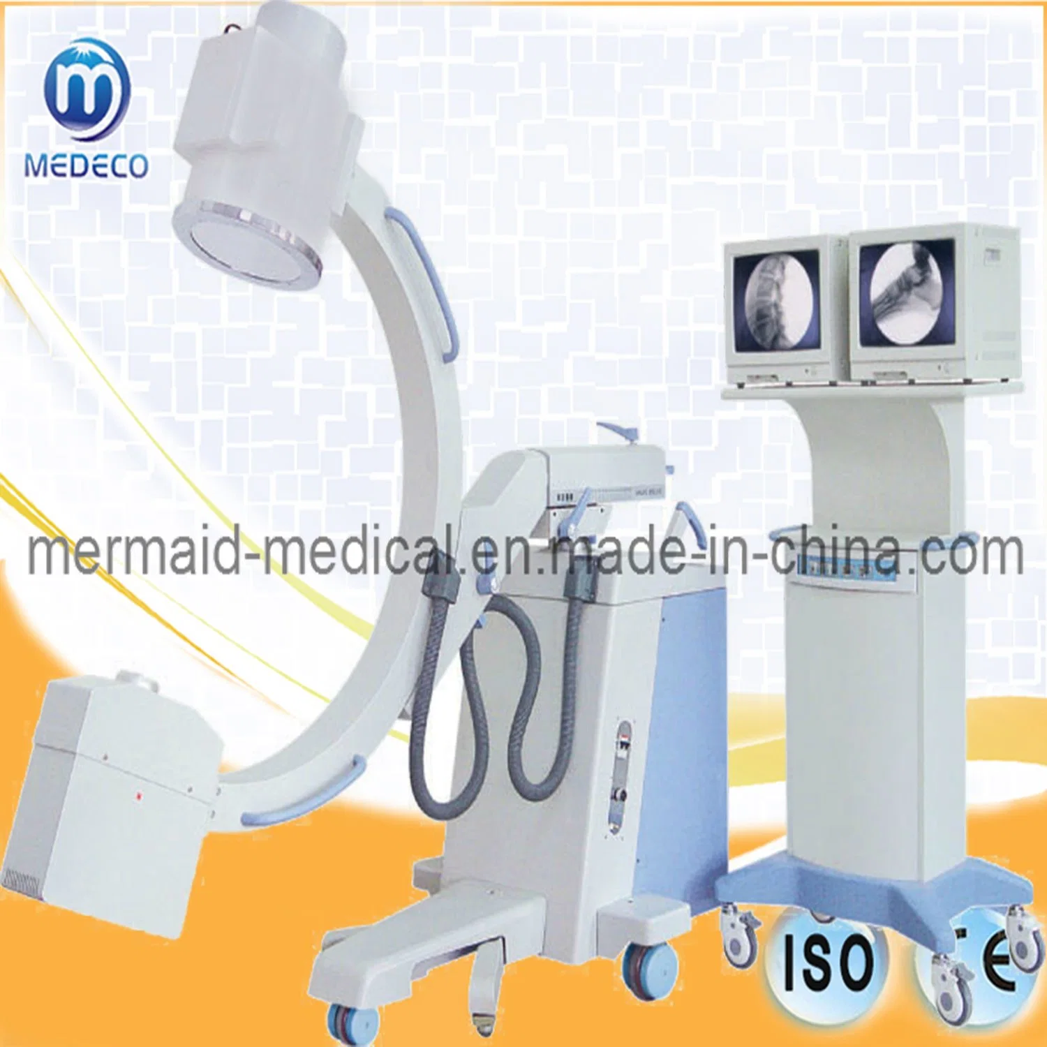
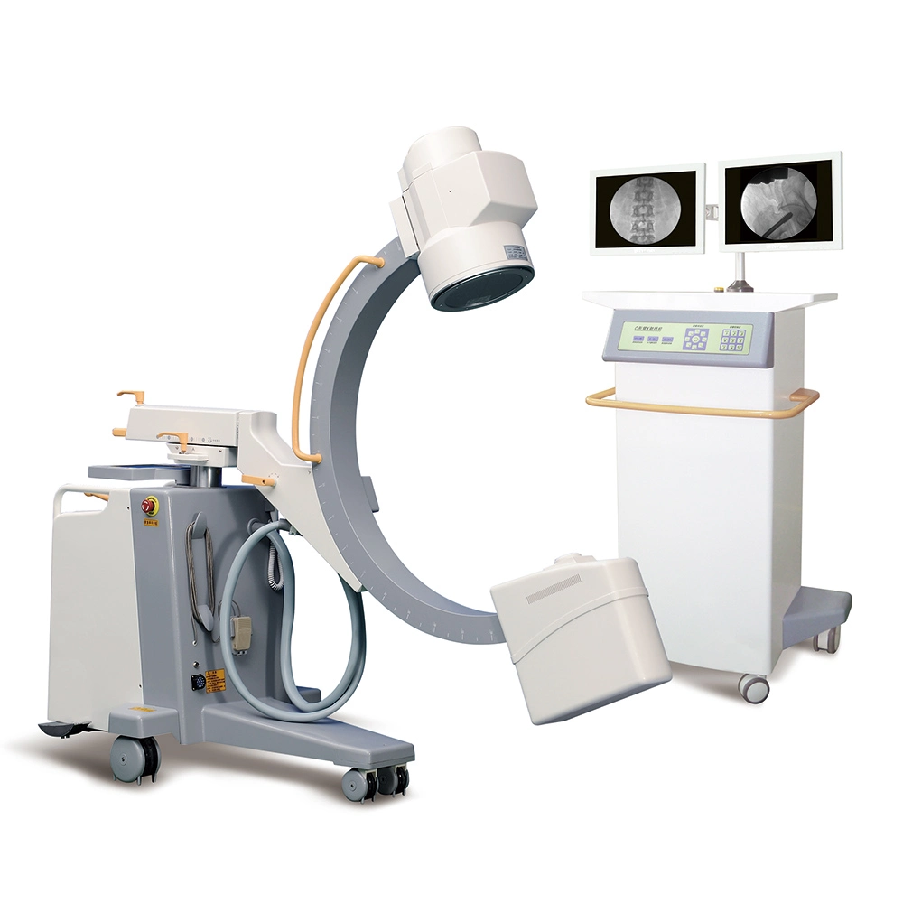
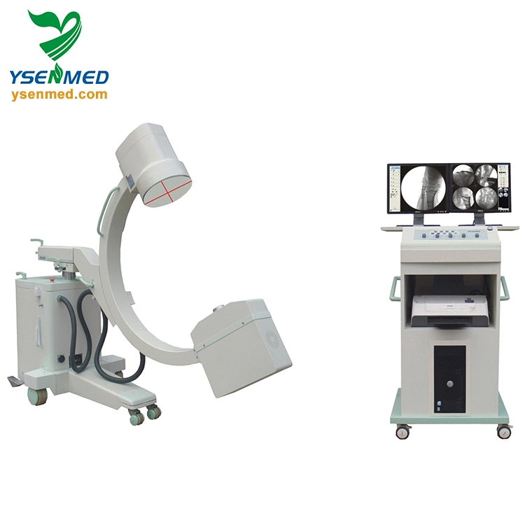
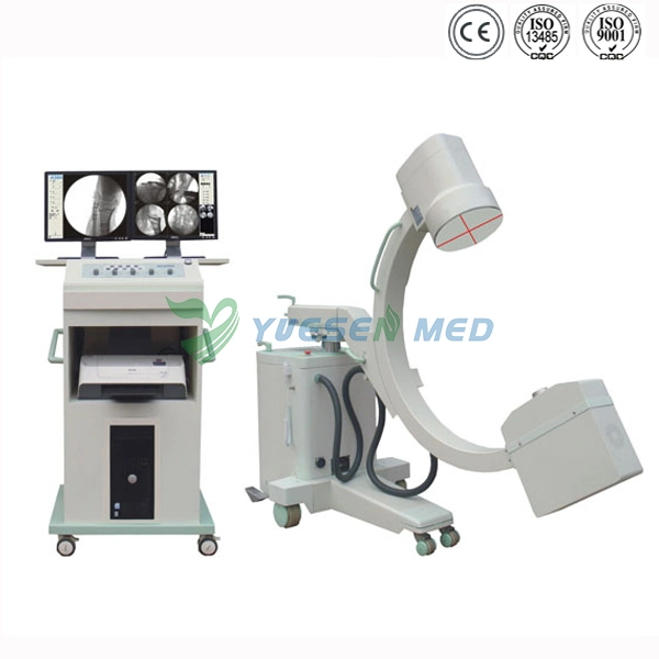
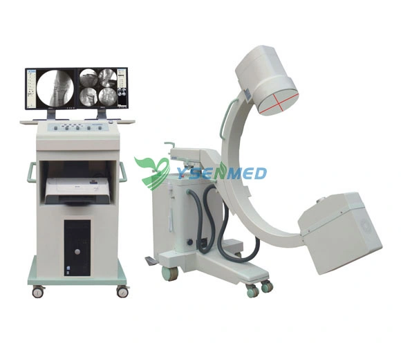
| C-arm machinery index | C-arm stander(with perfect C arm balance system) of ultra-quiet motor-driven up and down, easy and handy to operate. The machine can maintain balance with up and down of the C arm. | |
| Distance between focus and window>900mm, arc depth>650mm, horizontal moving range :200mm vertical moving range:400mm | |
| Tilt angle :±12.5° rotation :±180°corner:125°(-35°-- +90°) | |
| Laser Orientation | Laser positioning function can help to position precisely and reduce exposure times in the operation, reduce unnecessary radiation. | |
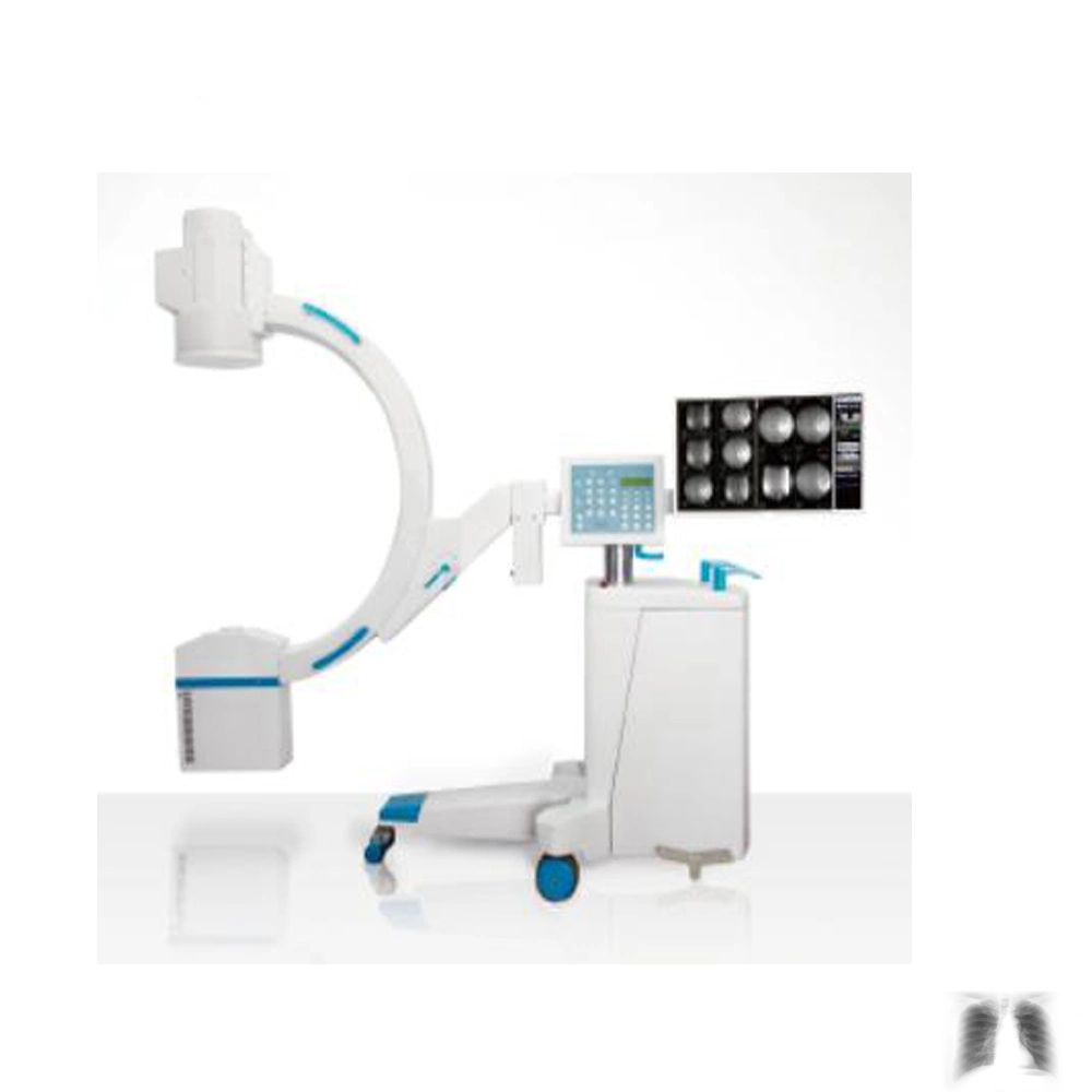
| Radiography mode | |
| Image intensifier & Real-time digital image(1.3 million pixel) processing system | |
| (CCD)Digital video camera | million pixels high-definition, high-speed CCD camera, a 1024 × 1024 matrix, 30 frames per second image acquisition, through real-time adjustment of window width, window level, noise reduction, sharpening and Gain adjustment to ensure high-quality and high-resolution images. | |
| Image resolution index | Grey level:10 Resolution:22 lp/cm | |
| Camera virtual rotation design | No X-ray needed, also called digital image rotation,display the right angle when exposure, superior to camera mechanical rotation | |
| Image processing system | 1.Real time edge enhancement(sharpening) | Image sharpening is also called edge enhancement, that is, to make blurry edge of the image clearer | |
| 2.Real time adjustment of the window width and window level | Obtain images by real-time adjustment of window width,window level, displaying images with different brightness. | |
| 3.Real time(static)multiform noise reduction | Noise reduction, and improve image clarity | |
| 4.Real tim GAIN adjustment | Can amplify image signal, reduce x-ray dose, suitable for patient with super large size. | |
| 5.Real tim dynamic brightness compensation and R calibration functions | Through dynamic logarithmic transformation,linear reduction the system signals and r calibration technology, dark parts of the image can be enhanced and clear rich-layer images can be obtained | |
| 6.Image conversing ,up-down and right-left rotating,and frame freezing | |
| 7.Patient information management ,Clinic report and print | |
| 8.The Image can be storage as JPG and DICOM 3.0 format, can be linked to hard-disk ,USB and DVD and printer,meet the need of sharing the network information ,communication and storage in hospital | |
| Display | Display2 17' TFT-LCD display | |
| Printer | Printer Laser / jet Printer | |
| Standard configuration | 1.Combined tube(5.0kW)IMD | 1 set; | |
| 2.Image intensifier (9 inch 3 fields of view) | 1 set | |
| 3.C-arm stander (with full C-arm balance system) | 1 set; | |
| 4.Exported grid | 1 set | |
| 5.Laser positioning | 1 set; | |
| 6.1.3million pixel 1024×1280×10bit camera | 1 set; | |
| 7.Electric adjustable beam limiting device | 1 set | |
| 8.17 inch LCD high resolution screen | 2 sets | |
| 9.Real-time digital imageprocession system (station) | 1 set; | |
| 10.Printer | 1 set | |
 Complaint