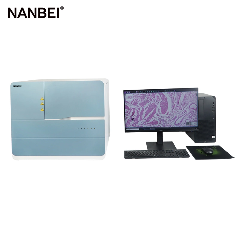
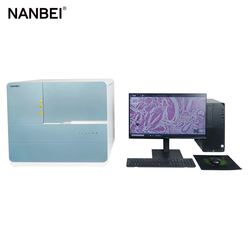
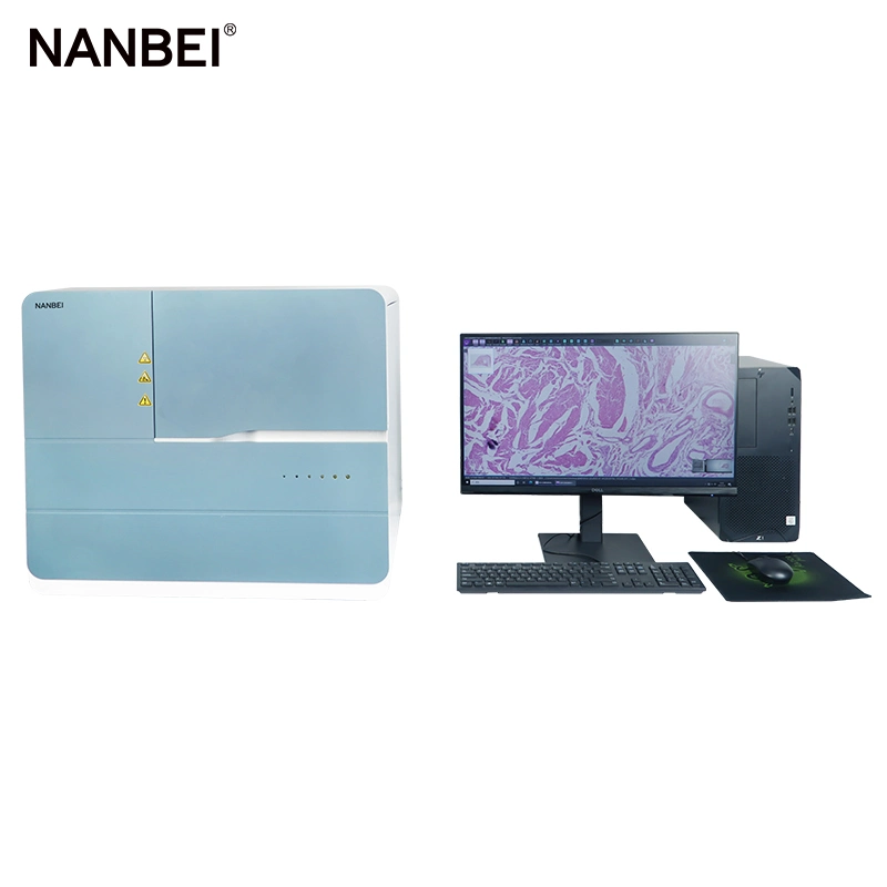
Description
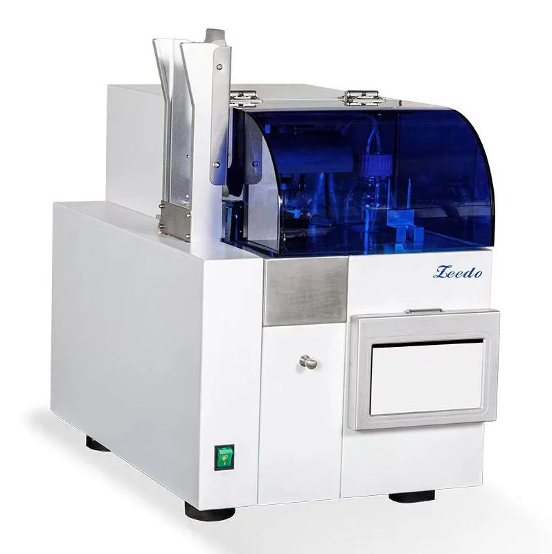
Fully Automatic Digital Pathology Slide Scanners
Digital Benefits
Digital slicing is the rapid scanning of pathological slide specimens into high-resolution electronic pictures, that is, the
process of rapid digitization of material slide specimens. Its essence is to achieve standardized, high-definition, and
full-information image acquisition;
Convenience for slice reading-make the film reading free from the limitation of the microscope, mouse operation can quickly select any part of the slice for observation, without being limited by the field of view under the microscope, with complete structure, comprehensive field of view, high resolution, clear pictures, saturated colors, and undistorted images ;
Slice preservation-analyze and classify similar pathological slice digital images through analysis and statistical software, making search and management more convenient, efficient and convenient. It avoids the breakage and discoloration of traditional slices in the process of production and use. Some precious specimens can be stored for a long time after being converted into digital slices, and they are not easy to lose.
Scientific research analysis-to ensure the consistency of image quality and the accuracy of each analysis data;Based on the usage habits of scientific research users, the software is adjusted, and the results are exported according to the requirements of scientific research articles and reports;
Slice preview information can be retrieved at any time, and panoramic result format conversion is supported, which facilitates the versatility of result analysis.Provide a complete secondary development interface, which can integrate artificial intelligence identification schemes in the instrument software system to realize functions such as digital diagnosis, intelligent identification, and automatic result interpretation.
| Product name | Fully Automatic Digital Slide Scanner |
| Model | NBScan-60 |
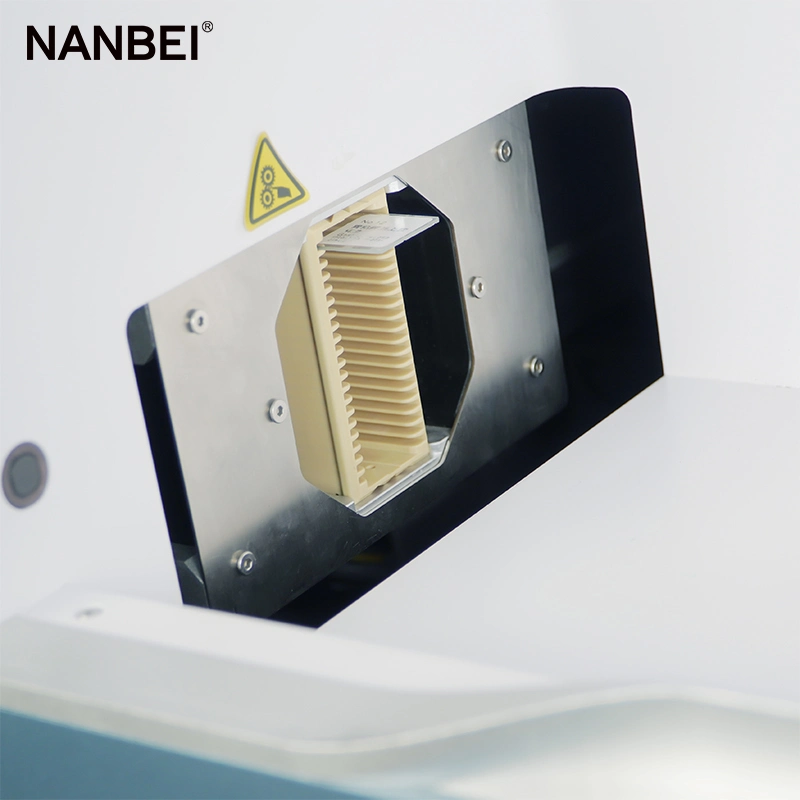
| Flux | 60 pieces |
| Objective lens | 20×, 40× double objective lens |
| Scan settings | Single-layer scanning, multi-layer scanning |
| Export area pictures | Supports 300 dpi printing resolution export, which can be used for reports or thesis illustrations |
| Dimensions | 635*595*571mm (scan host) |
| Weight | 60KG |
| Slice reading mode | Support local, multi-browser, other mobile terminal viewing (optional) |
| Voltage | 220V 50HZ |
| Place of Origin | China |
S-shaped track area scan
(scanning method)
Save scanning time and improve scanning accuracy
Compared with the Z-type unidirectional linear scanning of the linear camera, the area array scanning method of the large target
surface camera is better (example A surface scanning, B line scanning); CMOS, high-quality pixels, camera frame rate ≥ 43 frames
per second, guaranteed scanning continuity and minimum number of image stitching.
Panorama storage compression is smaller
High-speed, efficient and unattended Based on the extreme compression technology of the special database of artificial intelligence algorithm, the storage space and cost are greatly saved without affecting the image quality.
Merge multi-layer images into one clear image
Z-stack, depth of field extension, multi-layer fusion technology
When shooting thick tissue (about 20 μm) or uneven tissue, each focal plane can only image a part of the area, and cannot capture a complete image at the same time. After the sample is layered and imaged through the Z-stack imaging and depth of field expansion function, the multi-layer images are fused to automatically select the clearest area, thereby showing a complete image of all uneven tissues in one image.
Fast autofocus
Wide-range focal plane fitting, field curvature digital correction
Intelligently select the focus position according to the tissue situation, and fit the focal plane of the entire tissue according to the selected focus value to ensure that the image of each field of view is clear. At the same time, the system supports the user to customize the number of focus points according to the flatness of the tissue, so as to achieve the optimal focus of the sample.
Automatic recognition of slide type
H&E stained sections, immunohistochemistry, cytology sections Applicable to histopathology: tissue section, cytopathology: TCT exfoliated cell smear, etc.
Q1: Why Choose NANBEI ?
(1).Professional manufacturer with more than 13 years experience
(2).Exported to more than 97% Countries
(3).Turnkey Solution is no problem
Q2:OEM,ODM acceptable or not?
Absolutely Yes
Q3:What's kind of Payment terms for customer choosing?
T/T ,Western Union, Money Gram , Credit Card, Paypal , L/C ...
Q4:Can we visit your factory online?
Absolutely no problem
Q5:Can online video inspection before shipment?
Absolutely no problem
Q6: what's the MOQ ? Sample order is OK?
MOQ:1 set, sample order is no problem
Q7:What's kind of shipment for customer choosing?
Usually ship by sea, by air, by international express .
We can also provide reasonable solutions according to your transportation requirements
Q8:How to ensure product quality and after-sales service?
We have CE, ISO quality certificate, and SGS authentication.
After-sale service:
1. Warranty : 1 year
2. We supply free part for quality problem in warranty
3. Long life technical support and service

 Complaint