


 Complaint

2. Summary of Main Specifications |
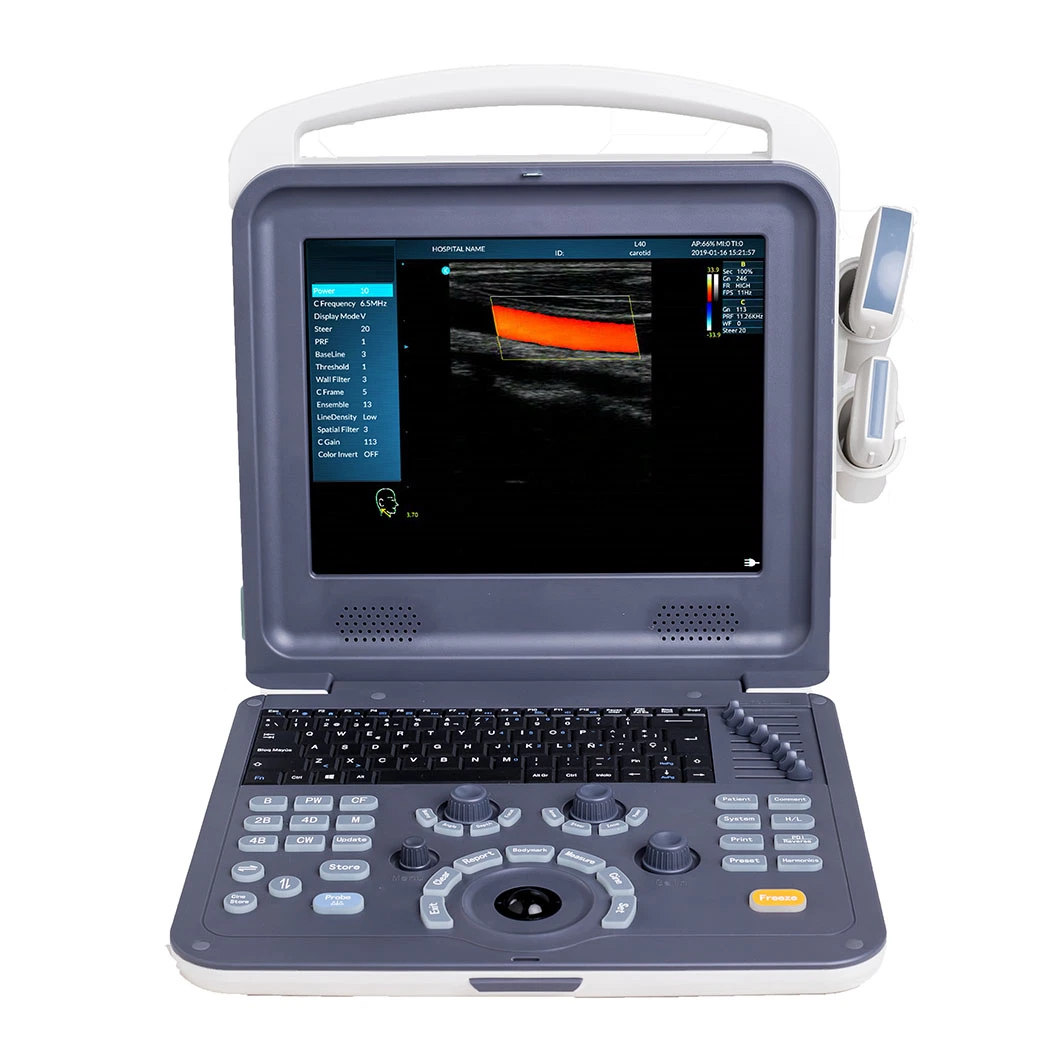
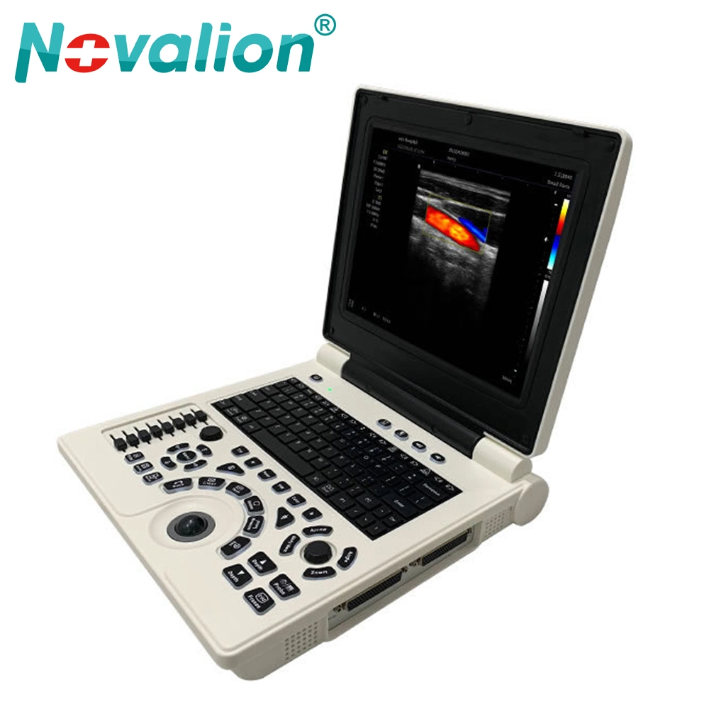
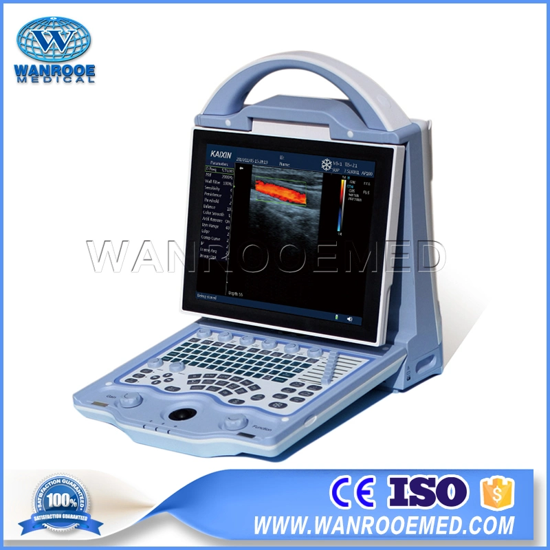
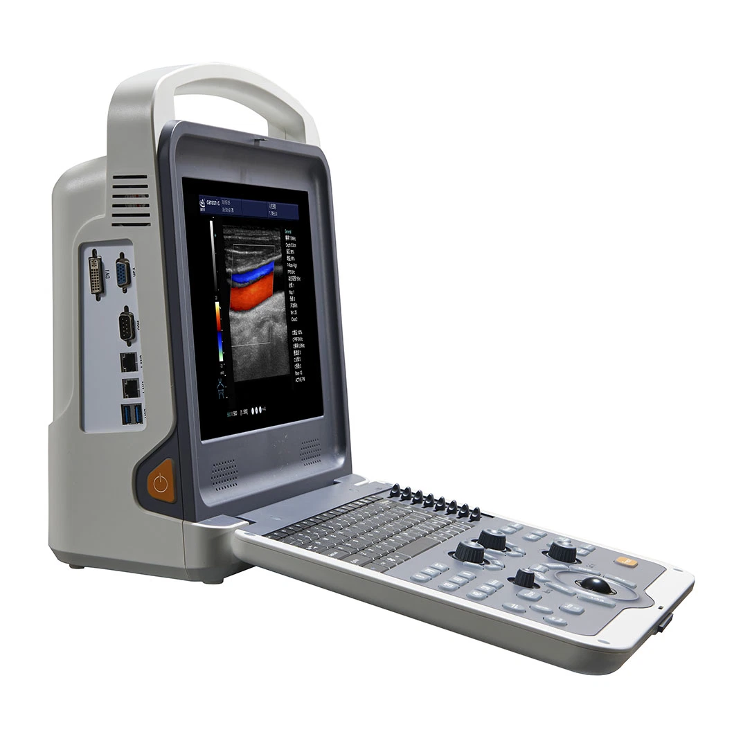
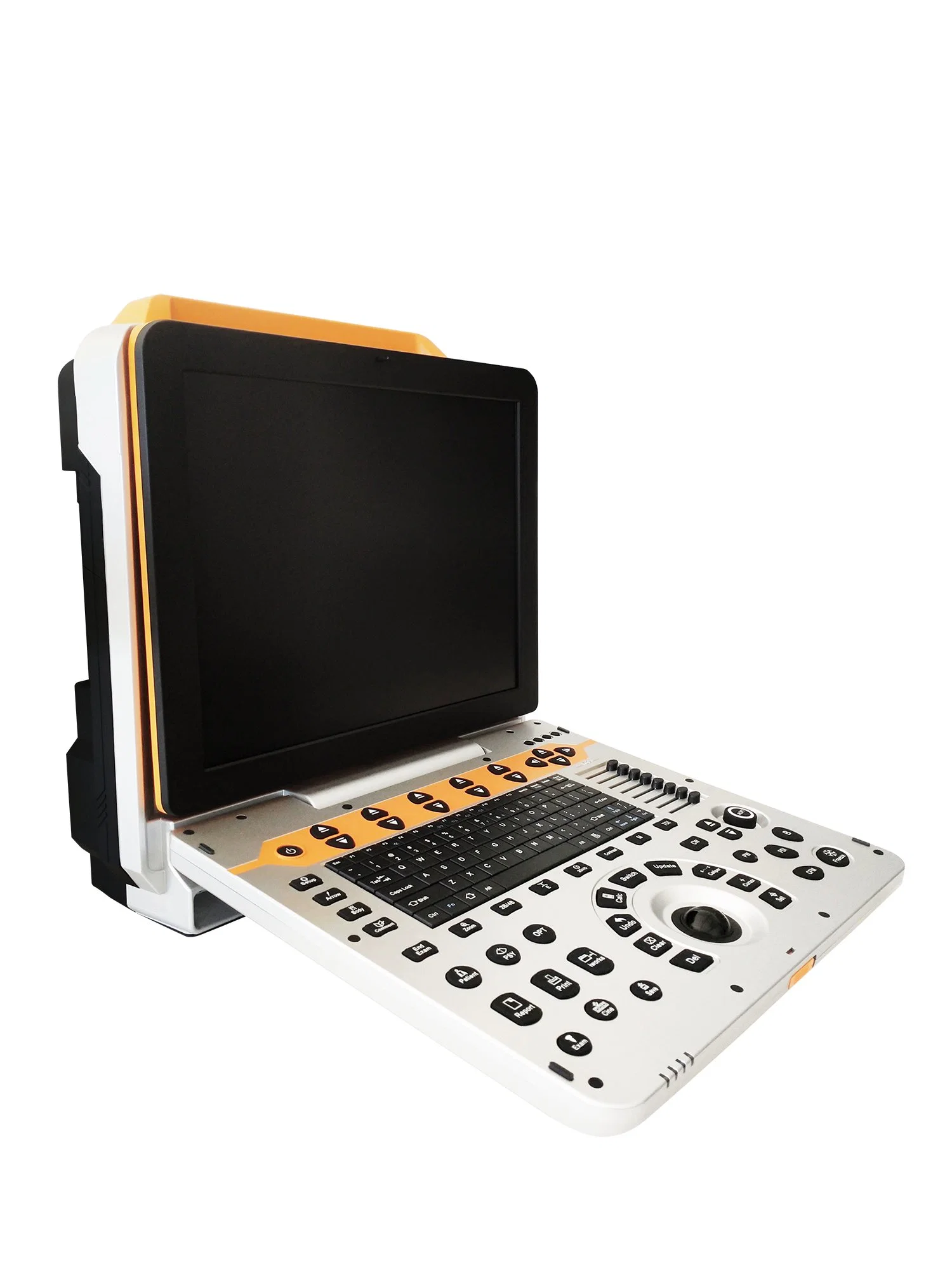
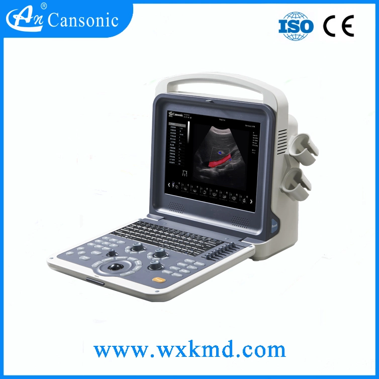
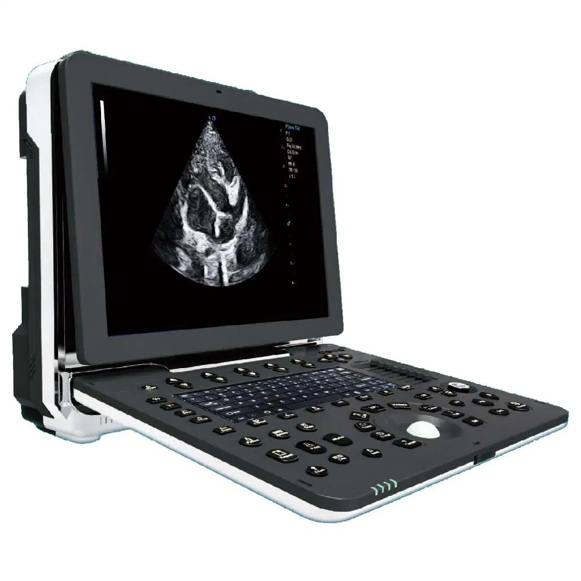
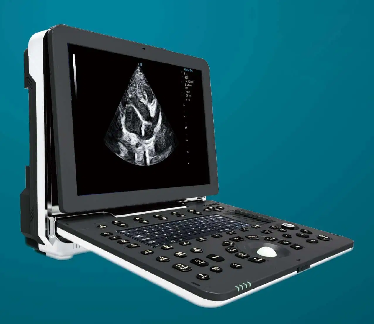
2. Summary of Main Specifications 3.1. Full Digital Color Doppler Ultrasonic Mainframe |
3.2. Full digital transmission and reception of beam synthesizer |
3.3. Multiple-beam Synthesis |
3.4. Full-Digital 2D Gray Scale Imaging |
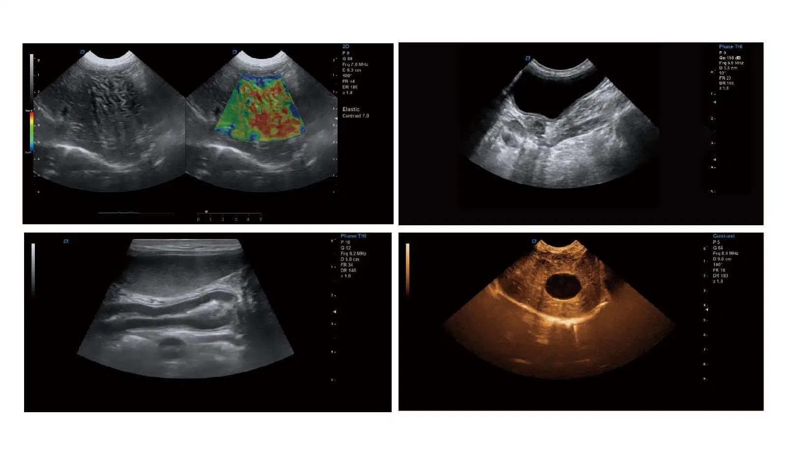
3.5. Tissue Harmonic Imaging (THI) |
3.6. B/C Real-timeTwo Synchronous Imaging |
3.7. M Mode Imaging |
3.8. Anatomic M Mode Imaging (Sampling line ≥3) |
3.9. Color Doppler Imaging (C, PDI, DPDI) |
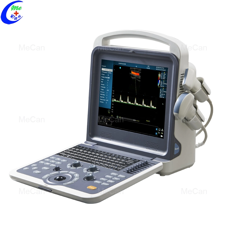
3.10. Spectral Doppler Imaging (PW, HPRF PW, CW) |
3.11. Tissue Doppler Imaging (TVI, M-mode, Spectral Imaging, etc.) |
3.12. Four-dimensional Supersonic Image Formation |
3.13.★Contrast Tuned Imaging |
3.14.★PView Wide Field Imaging |
3.15. Space Compound Imaging (application to Abdominal, GYN, vessel, superficial small organs, can be dual image contrast display at same time) |
3.16. Frequency & Focus Compound |
3.17. Extended field-of-view (EFOV) |
3.18. Real-time dual image contrast display |
3.19. B/C/D Real-time three synchronous imaging |
3.20. Speckle Reduce Imaging (SRI) |
3.21. Elastography |